[English] 日本語
Yorodumi
- EMDB-24827: CryoEM structure of Methylococcus capsulatus (Bath) pMMO in a nat... -

+
Open data


ID or keywords:

Loading...

-
Basic information

Entry
Database: EMDB / ID: EMD-24827
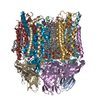
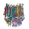
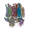
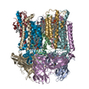
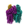
TitleCryoEM structure of Methylococcus capsulatus (Bath) pMMO in a native lipid nanodisc at 2.26 Angstrom resolution
Map dataCryoEM structure of Methylococcus capsulatus (Bath) pMMO in a POPC lipid nanodisc at 2.26 angstrom resolution
Sample
  • Complex: pMMO complex in a native lipid nanodisc
    • Protein or peptide: Particulate methane monooxygenase alpha subunit
    • Protein or peptide: Ammonia monooxygenase/methane monooxygenase, subunit C family protein
    • Protein or peptide: Particulate methane monooxygenase beta subunit
  • Ligand: COPPER (II) ION
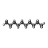
  • Ligand: DECANE
  • Ligand: DIUNDECYL PHOSPHATIDYL CHOLINE
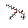
  • Ligand: 1,2-dihexanoyl-sn-glycero-3-phosphocholine
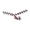
  • Ligand: 1,2-DIDECANOYL-SN-GLYCERO-3-PHOSPHOCHOLINE
  • Ligand: water
Function / homology
Function and homology information


methane monooxygenase (particulate) / methane monooxygenase complex / methane monooxygenase activity / methane monooxygenase (soluble) / methane monooxygenase NADH activity / methane monooxygenase NADPH activity / methane metabolic process / monooxygenase activity / membrane / metal ion binding
Similarity search - Function
Ammonia monooxygenase/particulate methane monooxygenase, subunit A / Ammonia monooxygenase/particulate methane monooxygenase, subunit C / Ammonia/methane monooxygenase, subunit B, hairpin domain superfamily / Ammonia/methane monooxygenase, subunit B, C-terminal / Ammonia monooxygenase/particulate methane monooxygenase, subunit C domain superfamily / Ammonia/particulate methane monooxygenase, subunit A superfamily / Ammonia monooxygenase / Ammonia monooxygenase/methane monooxygenase, subunit C / Ammonia monooxygenase/particulate methane monooxygenase, subunit B / Ammonia/methane monooxygenase, subunitB, N-terminal / Monooxygenase subunit B protein
Similarity search - Domain/homology
Particulate methane monooxygenase alpha subunit / Ammonia monooxygenase/methane monooxygenase, subunit C family protein / Particulate methane monooxygenase beta subunit
Similarity search - Component
Biological speciesMethylococcus capsulatus (strain ATCC 33009 / NCIMB 11132 / Bath) (bacteria) / Methylococcus capsulatus str. Bath (bacteria)
Methodsingle particle reconstruction / cryo EM / Resolution: 2.26 Å
AuthorsKoo CW / Rosenzweig AC
Funding support United States, 1 items
OrganizationGrant numberCountry
National Institutes of Health/National Institute of General Medical Sciences (NIH/NIGMS) United States
CitationJournal: Science / Year: 2022
Title: Recovery of particulate methane monooxygenase structure and activity in a lipid bilayer.
Authors: Christopher W Koo / Frank J Tucci / Yuan He / Amy C Rosenzweig /
Abstract: Bacterial methane oxidation using the enzyme particulate methane monooxygenase (pMMO) contributes to the removal of environmental methane, a potent greenhouse gas. Crystal structures determined using ...Bacterial methane oxidation using the enzyme particulate methane monooxygenase (pMMO) contributes to the removal of environmental methane, a potent greenhouse gas. Crystal structures determined using inactive, detergent-solubilized pMMO lack several conserved regions neighboring the proposed active site. We show that reconstituting pMMO in nanodiscs with lipids extracted from the native organism restores methane oxidation activity. Multiple nanodisc-embedded pMMO structures determined by cryo-electron microscopy to 2.14- to 2.46-angstrom resolution reveal the structure of pMMO in a lipid environment. The resulting model includes stabilizing lipids, regions of the PmoA and PmoC subunits not observed in prior structures, and a previously undetected copper-binding site in the PmoC subunit with an adjacent hydrophobic cavity. These structures provide a revised framework for understanding and engineering pMMO function.
History
DepositionSep 9, 2021-
Header (metadata) releaseMar 30, 2022-
Map releaseMar 30, 2022-
UpdateMar 30, 2022-
Current statusMar 30, 2022Processing site: RCSB / Status: Released

-
Structure visualization

Supplemental images

Downloads & links

-
Map

FileDownload / File: emd_24827.map.gz / Format: CCP4 / Size: 307.5 MB / Type: IMAGE STORED AS FLOATING POINT NUMBER (4 BYTES)
AnnotationCryoEM structure of Methylococcus capsulatus (Bath) pMMO in a POPC lipid nanodisc at 2.26 angstrom resolution
Voxel sizeX=Y=Z: 0.47222 Å
Density
Contour LevelBy AUTHOR: 0.0229
Minimum - Maximum-0.0441517 - 0.11982533
Average (Standard dev.)0.00020982897 (±0.005364079)
SymmetrySpace group: 1
Details

EMDB XML:

Map geometry
Axis orderXYZ
Origin000
Dimensions432432432
Spacing432432432
CellA=B=C: 204.0 Å
α=β=γ: 90.0 °

-
Supplemental data

-
Sample components

+
Entire : pMMO complex in a native lipid nanodisc

EntireName: pMMO complex in a native lipid nanodisc
Components
  • Complex: pMMO complex in a native lipid nanodisc
    • Protein or peptide: Particulate methane monooxygenase alpha subunit
    • Protein or peptide: Ammonia monooxygenase/methane monooxygenase, subunit C family protein
    • Protein or peptide: Particulate methane monooxygenase beta subunit
  • Ligand: COPPER (II) ION
  • Ligand: DECANE
  • Ligand: DIUNDECYL PHOSPHATIDYL CHOLINE
  • Ligand: 1,2-dihexanoyl-sn-glycero-3-phosphocholine
  • Ligand: 1,2-DIDECANOYL-SN-GLYCERO-3-PHOSPHOCHOLINE
  • Ligand: water

+
Supramolecule #1: pMMO complex in a native lipid nanodisc

SupramoleculeName: pMMO complex in a native lipid nanodisc / type: complex / ID: 1 / Parent: 0 / Macromolecule list: #1-#3
Source (natural)Organism: Methylococcus capsulatus (strain ATCC 33009 / NCIMB 11132 / Bath) (bacteria)

+
Macromolecule #1: Particulate methane monooxygenase alpha subunit

MacromoleculeName: Particulate methane monooxygenase alpha subunit / type: protein_or_peptide / ID: 1 / Number of copies: 3 / Enantiomer: LEVO / EC number: methane monooxygenase (particulate)
Source (natural)Organism: Methylococcus capsulatus str. Bath (bacteria) / Strain: ATCC 33009 / NCIMB 11132 / Bath
Molecular weightTheoretical: 46.129746 KDa
SequenceString: MKTIKDRIAK WSAIGLLSAV AATAFYAPSA SAHGEKSQAA FMRMRTIHWY DLSWSKEKVK INETVEIKGK FHVFEGWPET VDEPDVAFL NVGMPGPVFI RKESYIGGQL VPRSVRLEIG KTYDFRVVLK ARRPGDWHVH TMMNVQGGGP IIGPGKWITV E GSMSEFRN ...String:
MKTIKDRIAK WSAIGLLSAV AATAFYAPSA SAHGEKSQAA FMRMRTIHWY DLSWSKEKVK INETVEIKGK FHVFEGWPET VDEPDVAFL NVGMPGPVFI RKESYIGGQL VPRSVRLEIG KTYDFRVVLK ARRPGDWHVH TMMNVQGGGP IIGPGKWITV E GSMSEFRN PVTTLTGQTV DLENYNEGNT YFWHAFWFAI GVAWIGYWSR RPIFIPRLLM VDAGRADELV SATDRKVAMG FL AATILIV VMAMSSANSK YPITIPLQAG TMRGMKPLEL PAPTVSVKVE DATYRVPGRA MRMKLTITNH GNSPIRLGEF YTA SVRFLD SDVYKDTTGY PEDLLAEDGL SVSDNSPLAP GETRTVDVTA SDAAWEVYRL SDIIYDPDSR FAGLLFFFDA TGNR QVVQI DAPLIPSFM

+
Macromolecule #2: Ammonia monooxygenase/methane monooxygenase, subunit C family protein

MacromoleculeName: Ammonia monooxygenase/methane monooxygenase, subunit C family protein
type: protein_or_peptide / ID: 2 / Number of copies: 3 / Enantiomer: LEVO / EC number: methane monooxygenase (soluble)
Source (natural)Organism: Methylococcus capsulatus str. Bath (bacteria) / Strain: ATCC 33009 / NCIMB 11132 / Bath
Molecular weightTheoretical: 29.839309 KDa
SequenceString: MAATTIGGAA AAEAPLLDKK WLTFALAIYT VFYLWVRWYE GVYGWSAGLD SFAPEFETYW MNFLYTEIVL EIVTASILWG YLWKTRDRN LAALTPREEL RRNFTHLVWL VAYAWAIYWG ASYFTEQDGT WHQTIVRDTD FTPSHIIEFY LSYPIYIITG F AAFIYAKT ...String:
MAATTIGGAA AAEAPLLDKK WLTFALAIYT VFYLWVRWYE GVYGWSAGLD SFAPEFETYW MNFLYTEIVL EIVTASILWG YLWKTRDRN LAALTPREEL RRNFTHLVWL VAYAWAIYWG ASYFTEQDGT WHQTIVRDTD FTPSHIIEFY LSYPIYIITG F AAFIYAKT RLPFFAKGIS LPYLVLVVGP FMILPNVGLN EWGHTFWFME ELFVAPLHYG FVIFGWLALA VMGTLTQTFY SF AQGGLGQ SLCEAVDEGL IAK

+
Macromolecule #3: Particulate methane monooxygenase beta subunit

MacromoleculeName: Particulate methane monooxygenase beta subunit / type: protein_or_peptide / ID: 3 / Number of copies: 3 / Enantiomer: LEVO / EC number: methane monooxygenase (particulate)
Source (natural)Organism: Methylococcus capsulatus str. Bath (bacteria) / Strain: ATCC 33009 / NCIMB 11132 / Bath
Molecular weightTheoretical: 28.445098 KDa
SequenceString: MSAAQSAVRS HAEAVQVSRT IDWMALFVVF FVIVGSYHIH AMLTMGDWDF WSDWKDRRLW VTVTPIVLVT FPAAVQSYLW ERYRLPWGA TVCVLGLLLG EWINRYFNFW GWTYFPINFV FPASLVPGAI ILDTVLMLSG SYLFTAIVGA MGWGLIFYPG N WPIIAPLH ...String:
MSAAQSAVRS HAEAVQVSRT IDWMALFVVF FVIVGSYHIH AMLTMGDWDF WSDWKDRRLW VTVTPIVLVT FPAAVQSYLW ERYRLPWGA TVCVLGLLLG EWINRYFNFW GWTYFPINFV FPASLVPGAI ILDTVLMLSG SYLFTAIVGA MGWGLIFYPG N WPIIAPLH VPVEYNGMLM SIADIQGYNY VRTGTPEYIR MVEKGTLRTF GKDVAPVSAF FSAFMSILIY FMWHFIGRWF SN ERFLQST

+
Macromolecule #4: COPPER (II) ION

MacromoleculeName: COPPER (II) ION / type: ligand / ID: 4 / Number of copies: 9 / Formula: CU
Molecular weightTheoretical: 63.546 Da
Chemical component information

ChemComp-CU:
COPPER (II) ION / Copper

+
Macromolecule #5: DECANE

MacromoleculeName: DECANE / type: ligand / ID: 5 / Number of copies: 18 / Formula: D10
Molecular weightTheoretical: 142.282 Da
Chemical component information

ChemComp-D10:
DECANE / Decane

+
Macromolecule #6: DIUNDECYL PHOSPHATIDYL CHOLINE

MacromoleculeName: DIUNDECYL PHOSPHATIDYL CHOLINE / type: ligand / ID: 6 / Number of copies: 18 / Formula: PLC
Molecular weightTheoretical: 622.834 Da
Chemical component information

ChemComp-PLC:
DIUNDECYL PHOSPHATIDYL CHOLINE / phospholipid*YM

+
Macromolecule #7: 1,2-dihexanoyl-sn-glycero-3-phosphocholine

MacromoleculeName: 1,2-dihexanoyl-sn-glycero-3-phosphocholine / type: ligand / ID: 7 / Number of copies: 6 / Formula: HXG
Molecular weightTheoretical: 454.515 Da
Chemical component information

ChemComp-HXG:
1,2-dihexanoyl-sn-glycero-3-phosphocholine

+
Macromolecule #8: 1,2-DIDECANOYL-SN-GLYCERO-3-PHOSPHOCHOLINE

MacromoleculeName: 1,2-DIDECANOYL-SN-GLYCERO-3-PHOSPHOCHOLINE / type: ligand / ID: 8 / Number of copies: 12 / Formula: P1O
Molecular weightTheoretical: 566.728 Da
Chemical component information

ChemComp-P1O:
1,2-DIDECANOYL-SN-GLYCERO-3-PHOSPHOCHOLINE / DDPC, phospholipid*YM

+
Macromolecule #9: water

MacromoleculeName: water / type: ligand / ID: 9 / Number of copies: 355 / Formula: HOH
Molecular weightTheoretical: 18.015 Da
Chemical component information

ChemComp-HOH:
WATER / Water

-
Experimental details

-
Structure determination

Methodcryo EM
Processingsingle particle reconstruction
Aggregation stateparticle

-
Sample preparation

BufferpH: 7.3
VitrificationCryogen name: ETHANE

-
Electron microscopy

MicroscopeFEI TITAN KRIOS
Electron beamAcceleration voltage: 300 kV / Electron source: FIELD EMISSION GUN
Electron opticsIllumination mode: FLOOD BEAM / Imaging mode: BRIGHT FIELDBright-field microscopy
Image recordingFilm or detector model: GATAN K3 (6k x 4k) / Average electron dose: 52.0 e/Å2
Experimental equipment
Model: Titan Krios / Image courtesy: FEI Company

-
Image processing

Initial angle assignmentType: MAXIMUM LIKELIHOOD
Final angle assignmentType: MAXIMUM LIKELIHOOD
Final reconstructionResolution.type: BY AUTHOR / Resolution: 2.26 Å / Resolution method: FSC 0.143 CUT-OFF / Number images used: 1477719
FSC plot (resolution estimation)

+
About Yorodumi

-
News

-
Feb 9, 2022. New format data for meta-information of EMDB entries

New format data for meta-information of EMDB entries

  • Version 3 of the EMDB header file is now the official format.
  • The previous official version 1.9 will be removed from the archive.

Related info.:EMDB header

External links:wwPDB to switch to version 3 of the EMDB data model

-
Aug 12, 2020. Covid-19 info

Covid-19 info

URL: https://pdbj.org/emnavi/covid19.php

New page: Covid-19 featured information page in EM Navigator.

Related info.:Covid-19 info / Mar 5, 2020. Novel coronavirus structure data

+
Mar 5, 2020. Novel coronavirus structure data

Novel coronavirus structure data

Related info.:Yorodumi Speices / Aug 12, 2020. Covid-19 info

External links:COVID-19 featured content - PDBj / Molecule of the Month (242):Coronavirus Proteases

+
Jan 31, 2019. EMDB accession codes are about to change! (news from PDBe EMDB page)

EMDB accession codes are about to change! (news from PDBe EMDB page)

  • The allocation of 4 digits for EMDB accession codes will soon come to an end. Whilst these codes will remain in use, new EMDB accession codes will include an additional digit and will expand incrementally as the available range of codes is exhausted. The current 4-digit format prefixed with “EMD-” (i.e. EMD-XXXX) will advance to a 5-digit format (i.e. EMD-XXXXX), and so on. It is currently estimated that the 4-digit codes will be depleted around Spring 2019, at which point the 5-digit format will come into force.
  • The EM Navigator/Yorodumi systems omit the EMD- prefix.

Related info.:Q: What is EMD? / ID/Accession-code notation in Yorodumi/EM Navigator

External links:EMDB Accession Codes are Changing Soon! / Contact to PDBj

+
Jul 12, 2017. Major update of PDB

Major update of PDB

  • wwPDB released updated PDB data conforming to the new PDBx/mmCIF dictionary.
  • This is a major update changing the version number from 4 to 5, and with Remediation, in which all the entries are updated.
  • In this update, many items about electron microscopy experimental information are reorganized (e.g. em_software).
  • Now, EM Navigator and Yorodumi are based on the updated data.

External links:wwPDB Remediation / Enriched Model Files Conforming to OneDep Data Standards Now Available in the PDB FTP Archive

-
Yorodumi

Thousand views of thousand structures

  • Yorodumi is a browser for structure data from EMDB, PDB, SASBDB, etc.
  • This page is also the successor to EM Navigator detail page, and also detail information page/front-end page for Omokage search.
  • The word "yorodu" (or yorozu) is an old Japanese word meaning "ten thousand". "mi" (miru) is to see.

Related info.:EMDB / PDB / SASBDB / Comparison of 3 databanks / Yorodumi Search / Aug 31, 2016. New EM Navigator & Yorodumi / Yorodumi Papers / Jmol/JSmol / Function and homology information / Changes in new EM Navigator and Yorodumi

Read more