National Institutes of Health/National Institute of General Medical Sciences (NIH/NIGMS)
United States
Citation
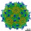
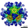
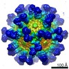
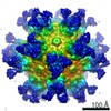
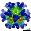
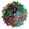
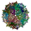
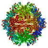
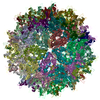
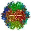
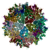
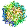
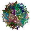
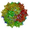
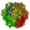
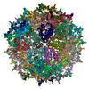
Journal: J Virol / Year: 2021 Title: Structural Study of Aavrh.10 Receptor and Antibody Interactions. Authors: Mario Mietzsch / Jennifer C Yu / Jane Hsi / Paul Chipman / Felix Broecker / Zhang Fuming / Robert J Linhardt / Peter H Seeberger / Regine Heilbronn / Robert McKenna / Mavis Agbandje-McKenna / Abstract: Recombinant adeno-associated virus (rAAV) vectors are one of the leading tools for the delivery of therapeutic genes in human gene therapy applications. For a successful transfer of their payload, ...Recombinant adeno-associated virus (rAAV) vectors are one of the leading tools for the delivery of therapeutic genes in human gene therapy applications. For a successful transfer of their payload, the AAV vectors have to circumvent potential preexisting neutralizing host antibodies and bind to the receptors of the target cells. Both of these aspects have not been structurally analyzed for AAVrh.10. Here, cryo-electron microscopy and three-dimensional image reconstruction were used to map the binding site of sulfated -acetyllactosamine (LacNAc; previously shown to bind AAVrh.10) and a series of four monoclonal antibodies (MAbs). LacNAc was found to bind to a pocket located on the side of the 3-fold capsid protrusion that is mostly conserved to AAV9 and equivalent to its galactose-binding site. As a result, AAVrh.10 was also shown to be able to bind to cell surface glycans with terminal galactose. For the antigenic characterization, it was observed that several anti-AAV8 MAbs cross-react with AAVrh.10. The binding sites of these antibodies were mapped to the 3-fold capsid protrusions. Based on these observations, the AAVrh.10 capsid surface was engineered to create variant capsids that escape these antibodies while maintaining infectivity. Gene therapy vectors based on adeno-associated virus rhesus isolate 10 (AAVrh.10) have been used in several clinical trials to treat monogenetic diseases. However, compared to other AAV serotypes little is known about receptor binding and antigenicity of the AAVrh.10 capsid. Particularly, preexisting neutralizing antibodies against capsids are an important challenge that can hamper treatment efficiency. This study addresses both topics and identifies critical regions of the AAVrh.10 capsid for receptor and antibody binding. The insights gained were utilized to generate AAVrh.10 variants capable of evading known neutralizing antibodies. The findings of this study could further aid the utilization of AAVrh.10 vectors in clinical trials and help the approval of the subsequent biologics.
#200 - Aug 2016 Quasisymmetry in Icosahedral Viruses similarity (1)
-
Assembly
Deposited unit
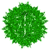
A: Capsid protein VP1 B: Capsid protein VP1 C: Capsid protein VP1 D: Capsid protein VP1 E: Capsid protein VP1 F: Capsid protein VP1 G: Capsid protein VP1 H: Capsid protein VP1 I: Capsid protein VP1 J: Capsid protein VP1 K: Capsid protein VP1 L: Capsid protein VP1 M: Capsid protein VP1 N: Capsid protein VP1 O: Capsid protein VP1 P: Capsid protein VP1 Q: Capsid protein VP1 R: Capsid protein VP1 S: Capsid protein VP1 T: Capsid protein VP1 U: Capsid protein VP1 V: Capsid protein VP1 W: Capsid protein VP1 X: Capsid protein VP1 Y: Capsid protein VP1 Z: Capsid protein VP1 a: Capsid protein VP1 b: Capsid protein VP1 c: Capsid protein VP1 d: Capsid protein VP1 e: Capsid protein VP1 f: Capsid protein VP1 g: Capsid protein VP1 h: Capsid protein VP1 i: Capsid protein VP1 j: Capsid protein VP1 k: Capsid protein VP1 l: Capsid protein VP1 m: Capsid protein VP1 n: Capsid protein VP1 o: Capsid protein VP1 p: Capsid protein VP1 q: Capsid protein VP1 r: Capsid protein VP1 s: Capsid protein VP1 t: Capsid protein VP1 u: Capsid protein VP1 v: Capsid protein VP1 w: Capsid protein VP1 x: Capsid protein VP1 y: Capsid protein VP1 z: Capsid protein VP1 1: Capsid protein VP1 2: Capsid protein VP1 3: Capsid protein VP1 4: Capsid protein VP1 5: Capsid protein VP1 6: Capsid protein VP1 7: Capsid protein VP1 8: Capsid protein VP1 hetero molecules
In the structure databanks used in Yorodumi, some data are registered as the other names, "COVID-19 virus" and "2019-nCoV". Here are the details of the virus and the list of structure data.
Jan 31, 2019. EMDB accession codes are about to change! (news from PDBe EMDB page)
EMDB accession codes are about to change! (news from PDBe EMDB page)
The allocation of 4 digits for EMDB accession codes will soon come to an end. Whilst these codes will remain in use, new EMDB accession codes will include an additional digit and will expand incrementally as the available range of codes is exhausted. The current 4-digit format prefixed with “EMD-” (i.e. EMD-XXXX) will advance to a 5-digit format (i.e. EMD-XXXXX), and so on. It is currently estimated that the 4-digit codes will be depleted around Spring 2019, at which point the 5-digit format will come into force.
The EM Navigator/Yorodumi systems omit the EMD- prefix.
Related info.:Q: What is EMD? / ID/Accession-code notation in Yorodumi/EM Navigator
Yorodumi is a browser for structure data from EMDB, PDB, SASBDB, etc.
This page is also the successor to EM Navigator detail page, and also detail information page/front-end page for Omokage search.
The word "yorodu" (or yorozu) is an old Japanese word meaning "ten thousand". "mi" (miru) is to see.
Related info.:EMDB / PDB / SASBDB / Comparison of 3 databanks / Yorodumi Search / Aug 31, 2016. New EM Navigator & Yorodumi / Yorodumi Papers / Jmol/JSmol / Function and homology information / Changes in new EM Navigator and Yorodumi
 Movie
Movie Controller


 Open data
 Basic information
 Components
 Keywords
 Function and homology information

 Authors
 United States, 1items
  Citation
 Structure visualization
 Downloads & links
 Other downloads
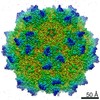


 PDBj
PDBj


 Assembly

 Type: D-saccharide, beta linking / Mass: 180.156 Da / Num. of mol.: 60 / Source method: obtained synthetically / Formula: C6H12O6 / Feature type: SUBJECT OF INVESTIGATION
 Sample preparation
 Electron microscopy imaging
 Processing