 Movie
Movie Controller

[English] 日本語
 Yorodumi
- PDB-7rwu: Crystal structure of Ni-bound RIDC1 variant in the presence of re... -
+ Open data

- Basic information
| Entry | Database: PDB / ID: 7rwu | ||||||||||||
|---|---|---|---|---|---|---|---|---|---|---|---|---|---|
| Title | Crystal structure of Ni-bound RIDC1 variant in the presence of reductant | ||||||||||||
 Components Components | Soluble cytochrome b562 | ||||||||||||
 Keywords Keywords | METAL BINDING PROTEIN | ||||||||||||
| Function / homology |  Function and homology information Function and homology information electron transport chain / electron transfer activity / periplasmic space / iron ion binding / heme binding electron transport chain / electron transfer activity / periplasmic space / iron ion binding / heme bindingSimilarity search - Function | ||||||||||||
| Biological species |  Escherichia coli (E. coli) Escherichia coli (E. coli) | ||||||||||||
| Method | X-RAY DIFFRACTION / SYNCHROTRON / MOLECULAR REPLACEMENT / Resolution: 1.8 Å | ||||||||||||
 Authors Authors | Kakkis, A. / Golub, E. | ||||||||||||
| Funding support |  United States, 3items United States, 3items
| ||||||||||||
 Citation Citation | Journal: Chem.Commun.(Camb.) / Year: 2022 Title: Redox- and metal-directed structural diversification in designed metalloprotein assemblies. Authors: Kakkis, A. / Golub, E. / Choi, T.S. / Tezcan, F.A. | ||||||||||||
| History |
|
- Structure visualization
| Structure viewer | Molecule: MolmilJmol/JSmol |
|---|
- Downloads & links
-Download
| PDBx/mmCIF format | 7rwu.cif.gz | 52.5 KB | Display | PDBx/mmCIF format |
|---|---|---|---|---|
| PDB format | pdb7rwu.ent.gz | 29.8 KB | Display | PDB format |
| PDBx/mmJSON format | 7rwu.json.gz | Tree view | PDBx/mmJSON format | |
| Others |  Other downloads Other downloads |
-Validation report
| Arichive directory | https://data.pdbj.org/pub/pdb/validation_reports/rw/7rwuftp://data.pdbj.org/pub/pdb/validation_reports/rw/7rwu | HTTPS FTP |
|---|
-Related structure data
| Related structure data | 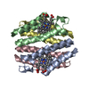 7rwvC 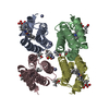 7rwwC 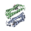 7rwxC 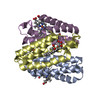 7rwyC 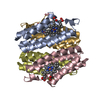 7su2C 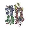 7tepC 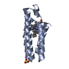 2bc5S S: Starting model for refinement C: citing same article ( |
|---|---|
| Similar structure data |
-Links
 PDBj
PDBj


- Assembly
| Deposited unit | 
| ||||||||||||||||||
|---|---|---|---|---|---|---|---|---|---|---|---|---|---|---|---|---|---|---|---|
| 1 | 
| ||||||||||||||||||
| Unit cell |
| ||||||||||||||||||
| Components on special symmetry positions |
|
-Components
-Protein , 1 types, 1 molecules A
| #1: Protein | Mass: 11657.113 Da / Num. of mol.: 1 Mutation: R56A, L60A, Q63W, K64S, K81H, D88W, V91I, D95H, D96A, K99H, T118C, R120C, Y123C Source method: isolated from a genetically manipulated source Source: (gene. exp.) Escherichia coli (E. coli) / Gene: cybC / Production host: Escherichia coli (E. coli) / References: UniProt: P0ABE7 |
|---|
-Non-polymers , 5 types, 179 molecules 


| #2: Chemical | Nickel Mass: 58.693 Da / Num. of mol.: 3 / Source method: obtained synthetically / Formula: Ni / Feature type: SUBJECT OF INVESTIGATION Mass: 58.693 Da / Num. of mol.: 3 / Source method: obtained synthetically / Formula: Ni / Feature type: SUBJECT OF INVESTIGATION#3: Chemical | ChemComp-TRS / | Tris Mass: 122.143 Da / Num. of mol.: 1 / Source method: obtained synthetically / Formula: C4H12NO3 / Comment: pH buffer*YM Mass: 122.143 Da / Num. of mol.: 1 / Source method: obtained synthetically / Formula: C4H12NO3 / Comment: pH buffer*YM#4: Chemical | ChemComp-HEC / | Heme C Mass: 618.503 Da / Num. of mol.: 1 / Source method: obtained synthetically / Formula: C34H34FeN4O4 Mass: 618.503 Da / Num. of mol.: 1 / Source method: obtained synthetically / Formula: C34H34FeN4O4#5: Chemical | ChemComp-CL / | Chloride Mass: 35.453 Da / Num. of mol.: 1 / Source method: obtained synthetically / Formula: Cl Mass: 35.453 Da / Num. of mol.: 1 / Source method: obtained synthetically / Formula: Cl#6: Water | ChemComp-HOH / | WaterMass: 18.015 Da / Num. of mol.: 173 / Source method: isolated from a natural source / Formula: H2O |
|---|
-Details
| Has ligand of interest | Y |
|---|
-Experimental details
-Experiment
| Experiment | Method: X-RAY DIFFRACTION / Number of used crystals: 1 |
|---|
- Sample preparation
| Crystal | Density Matthews: 2.99 Å3/Da / Density % sol: 58.91 % |
|---|---|
| Crystal grow | Temperature: 295 K / Method: vapor diffusion, sitting drop / pH: 8.5 / Details: 45% MPD, 0.1 M Tris, 0.2 M MgCl2 |
-Data collection
| Diffraction | Mean temperature: 80 K / Serial crystal experiment: N |
|---|---|
| Diffraction source | Source: SYNCHROTRON / Site: SSRL / Beamline: BL9-2 / Wavelength: 0.97946 Å |
| Detector | Type: DECTRIS PILATUS 6M / Detector: PIXEL / Date: Jul 21, 2021 |
| Radiation | Protocol: SINGLE WAVELENGTH / Monochromatic (M) / Laue (L): M / Scattering type: x-ray |
| Radiation wavelength | Wavelength: 0.97946 Å / Relative weight: 1 |
| Reflection | Resolution: 1.8→42.15 Å / Num. obs: 25101 / % possible obs: 99.99 % / Redundancy: 68 % / Biso Wilson estimate: 18.96 Å2 / CC1/2: 1 / Rmerge(I) obs: 0.069 / Rpim(I) all: 0.011 / Rrim(I) all: 0.07 / Net I/σ(I): 58.1 |
| Reflection shell | Resolution: 1.8→1.8 Å / Rmerge(I) obs: 0.191 / Num. unique obs: 1344 / CC1/2: 0.998 / Rpim(I) all: 0.032 / Rrim(I) all: 0.193 |
- Processing
| Software |
| ||||||||||||||||||||||||||||||||||||||||||||||||||||||||||||||||||||||
|---|---|---|---|---|---|---|---|---|---|---|---|---|---|---|---|---|---|---|---|---|---|---|---|---|---|---|---|---|---|---|---|---|---|---|---|---|---|---|---|---|---|---|---|---|---|---|---|---|---|---|---|---|---|---|---|---|---|---|---|---|---|---|---|---|---|---|---|---|---|---|---|
| Refinement | Method to determine structure: MOLECULAR REPLACEMENT Starting model: 2bc5 Resolution: 1.8→42.15 Å / SU ML: 0.1797 / Cross valid method: FREE R-VALUE / σ(F): 1.42 / Phase error: 18.8368 Stereochemistry target values: GeoStd + Monomer Library + CDL v1.2
| ||||||||||||||||||||||||||||||||||||||||||||||||||||||||||||||||||||||
| Solvent computation | Shrinkage radii: 0.9 Å / VDW probe radii: 1.11 Å / Solvent model: FLAT BULK SOLVENT MODEL | ||||||||||||||||||||||||||||||||||||||||||||||||||||||||||||||||||||||
| Displacement parameters | Biso mean: 23.6 Å2 | ||||||||||||||||||||||||||||||||||||||||||||||||||||||||||||||||||||||
| Refinement step | Cycle: LAST / Resolution: 1.8→42.15 Å
| ||||||||||||||||||||||||||||||||||||||||||||||||||||||||||||||||||||||
| Refine LS restraints |
| ||||||||||||||||||||||||||||||||||||||||||||||||||||||||||||||||||||||
| LS refinement shell |
|
