 Movie
Movie Controller


+ Open data

- Basic information
| Entry | Database: PDB / ID: 7qvk | ||||||
|---|---|---|---|---|---|---|---|
| Title | NM-02 in complex with HER2-ECD | ||||||
 Components Components |
| ||||||
 Keywords Keywords |  ONCOPROTEIN / receptor tyrosine protein kinase ONCOPROTEIN / receptor tyrosine protein kinase | ||||||
| Function / homology |  Function and homology information Function and homology informationnegative regulation of immature T cell proliferation in thymus / ERBB3:ERBB2 complex / ERBB2-ERBB4 signaling pathway / GRB7 events in ERBB2 signaling / immature T cell proliferation in thymus / RNA polymerase I core binding / regulation of microtubule-based process / ErbB-3 class receptor binding / semaphorin receptor complex / Sema4D induced cell migration and growth-cone collapse ...negative regulation of immature T cell proliferation in thymus / ERBB3:ERBB2 complex / ERBB2-ERBB4 signaling pathway / GRB7 events in ERBB2 signaling / immature T cell proliferation in thymus / RNA polymerase I core binding / regulation of microtubule-based process / ErbB-3 class receptor binding / semaphorin receptor complex / Sema4D induced cell migration and growth-cone collapse / motor neuron axon guidance / neurotransmitter receptor localization to postsynaptic specialization membrane / PLCG1 events in ERBB2 signaling / ERBB2-EGFR signaling pathway / neuromuscular junction development / positive regulation of Rho protein signal transduction / ERBB2 Activates PTK6 Signaling / Drug-mediated inhibition of ERBB2 signaling / Resistance of ERBB2 KD mutants to trastuzumab / Resistance of ERBB2 KD mutants to sapitinib / Resistance of ERBB2 KD mutants to tesevatinib / Resistance of ERBB2 KD mutants to neratinib / Resistance of ERBB2 KD mutants to osimertinib / Resistance of ERBB2 KD mutants to afatinib / Resistance of ERBB2 KD mutants to AEE788 / Resistance of ERBB2 KD mutants to lapatinib / Drug resistance in ERBB2 TMD/JMD mutants / enzyme-linked receptor protein signaling pathway / positive regulation of transcription by RNA polymerase I / ERBB2-ERBB3 signaling pathway / oligodendrocyte differentiation / ERBB2 Regulates Cell Motility / semaphorin-plexin signaling pathway / PI3K events in ERBB2 signaling / positive regulation of cell adhesion / positive regulation of protein targeting to membrane / regulation of angiogenesis / coreceptor activity / Schwann cell development / Signaling by ERBB2 / cellular response to epidermal growth factor stimulus / myelination / Downregulation of ERBB2:ERBB3 signaling / GRB2 events in ERBB2 signaling / TFAP2 (AP-2) family regulates transcription of growth factors and their receptors / transmembrane receptor protein tyrosine kinase activity / SHC1 events in ERBB2 signaling / Constitutive Signaling by Overexpressed ERBB2 / neurogenesis / basal plasma membrane / regulation of ERK1 and ERK2 cascade / phosphatidylinositol 3-kinase/protein kinase B signal transduction / positive regulation of translation / positive regulation of epithelial cell proliferation / cell surface receptor protein tyrosine kinase signaling pathway / Signaling by ERBB2 TMD/JMD mutants / positive regulation of MAP kinase activity / wound healing / neuromuscular junction / Signaling by ERBB2 ECD mutants / neuron differentiation / Signaling by ERBB2 KD Mutants / receptor protein-tyrosine kinase / receptor tyrosine kinase binding / cellular response to growth factor stimulus / Downregulation of ERBB2 signaling / ruffle membrane / peptidyl-tyrosine phosphorylation / Constitutive Signaling by Aberrant PI3K in Cancer / transmembrane signaling receptor activity / PIP3 activates AKT signaling / myelin sheath / presynaptic membrane / heart development / PI5P, PP2A and IER3 Regulate PI3K/AKT Signaling / RAF/MAP kinase cascade / positive regulation of cell growth / basolateral plasma membrane / protein tyrosine kinase activity / positive regulation of MAPK cascade / early endosome / cell surface receptor signaling pathway / receptor complex / endosome membrane / intracellular signal transduction / apical plasma membrane / positive regulation of protein phosphorylation / protein heterodimerization activity / protein phosphorylation / signaling receptor binding / positive regulation of cell population proliferation / negative regulation of apoptotic process / perinuclear region of cytoplasm / signal transduction / nucleoplasm / ATP binding / membrane / identical protein binding / nucleus / plasma membraneSimilarity search - Function | ||||||
| Biological species |  Homo sapiens (human) Homo sapiens (human) Camelus bactrianus (Bactrian camel) Camelus bactrianus (Bactrian camel) | ||||||
| Method | X-RAY DIFFRACTION / SYNCHROTRON / MOLECULAR REPLACEMENT / Resolution: 3.1 Å | ||||||
 Authors Authors | Cowan, R. / Hall, G. / Carr, M. | ||||||
| Funding support | 1items
| ||||||
 Citation Citation | Journal: Plos One / Year: 2023 Title: Co-crystallisation and humanisation of an anti-HER2 single-domain antibody as a theranostic tool. Authors: Sawmynaden, K. / Wong, N. / Davies, S. / Cowan, R. / Brown, R. / Tang, D. / Henry, M. / Tickle, D. / Matthews, D. / Carr, M. / Bakrania, P. / Hoi Ting, H. / Hall, G. | ||||||
| History |
|
- Structure visualization
| Structure viewer | Molecule: MolmilJmol/JSmol |
|---|
- Downloads & links
-Download
| PDBx/mmCIF format | 7qvk.cif.gz | 235.3 KB | Display | PDBx/mmCIF format |
|---|---|---|---|---|
| PDB format | pdb7qvk.ent.gz | Display | PDB format | |
| PDBx/mmJSON format | 7qvk.json.gz | Tree view | PDBx/mmJSON format | |
| Others |  Other downloads Other downloads |
-Validation report
| Arichive directory | https://data.pdbj.org/pub/pdb/validation_reports/qv/7qvkftp://data.pdbj.org/pub/pdb/validation_reports/qv/7qvk | HTTPS FTP |
|---|
-Related structure data
| Related structure data | 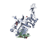 5my6S S: Starting model for refinement |
|---|---|
| Similar structure data |
-Links
 PDBj
PDBj


- Assembly
| Deposited unit | 
| ||||||||
|---|---|---|---|---|---|---|---|---|---|
| 1 |
| ||||||||
| Unit cell |
|
-Components
| #1: Protein | Mass: 69622.977 Da / Num. of mol.: 1 Source method: isolated from a genetically manipulated source Source: (gene. exp.) Homo sapiens (human) / Gene: ERBB2, HER2, MLN19, NEU, NGL / Cell line (production host): HEK293F / Production host: Homo sapiens (human)References: UniProt: P04626, receptor protein-tyrosine kinase |
|---|---|
| #2: Antibody | Mass: 14334.452 Da / Num. of mol.: 1 Source method: isolated from a genetically manipulated source Source: (gene. exp.) Camelus bactrianus (Bactrian camel) / Cell line (production host): ExpiCHO-S / Production host:  Cricetulus griseus (Chinese hamster) Cricetulus griseus (Chinese hamster) |
| #3: Polysaccharide | beta-D-mannopyranose-(1-4)-2-acetamido-2-deoxy-beta-D-glucopyranose-(1-4)-2-acetamido-2-deoxy-beta- ...beta-D-mannopyranose-(1-4)-2-acetamido-2-deoxy-beta-D-glucopyranose-(1-4)-2-acetamido-2-deoxy-beta-D-glucopyranose / Mass: 586.542 Da / Num. of mol.: 1 Source method: isolated from a genetically manipulated source |
| #4: Sugar | ChemComp-NAG / N-Acetylglucosamine  Type: D-saccharide, beta linking / Mass: 221.208 Da / Num. of mol.: 1 / Source method: obtained synthetically / Formula: C8H15NO6 Type: D-saccharide, beta linking / Mass: 221.208 Da / Num. of mol.: 1 / Source method: obtained synthetically / Formula: C8H15NO6 |
| Has ligand of interest | N |
-Experimental details
-Experiment
| Experiment | Method: X-RAY DIFFRACTION / Number of used crystals: 1 |
|---|
- Sample preparation
| Crystal | Density Matthews: 3.578768 Å3/Da / Density % sol: 65.65199 % |
|---|---|
| Crystal grow | Temperature: 291 K / Method: vapor diffusion, hanging drop / pH: 6.5 Details: 13% PEG6000 (w/v), 0.1 M MES pH 6.5, 7% 2-methyl-2,4-pentanediol |
-Data collection
| Diffraction | Mean temperature: 100 K / Serial crystal experiment: N |
|---|---|
| Diffraction source | Source: SYNCHROTRON / Site: Diamond  / Beamline: I24 / Wavelength: 0.96872 Å / Beamline: I24 / Wavelength: 0.96872 Å |
| Detector | Type: DECTRIS PILATUS3 6M / Detector: PIXEL / Date: Jun 26, 2019 |
| Radiation | Protocol: SINGLE WAVELENGTH / Monochromatic (M) / Laue (L): M / Scattering type: x-ray |
| Radiation wavelength | Wavelength: 0.96872 Å / Relative weight: 1 |
| Reflection | Resolution: 3.1→65.324 Å / Num. obs: 22490 / % possible obs: 100 % / Redundancy: 19.2 % / Biso Wilson estimate: 101.26 Å2 / CC1/2: 0.99 / Net I/σ(I): 5.1 |
| Reflection shell | Resolution: 3.1→3.31 Å / Redundancy: 19.7 % / Mean I/σ(I) obs: 0.7 / Num. unique obs: 4001 / CC1/2: 0.34 / % possible all: 100 |
- Processing
| Software |
| ||||||||||||||||||||||||||||||||||||||||||||||||||||||||||||||||||||||||||||||||||||||||||||||||||||||||||||||||||||||||||||||||||||||||||||||||||||||||||||||||
|---|---|---|---|---|---|---|---|---|---|---|---|---|---|---|---|---|---|---|---|---|---|---|---|---|---|---|---|---|---|---|---|---|---|---|---|---|---|---|---|---|---|---|---|---|---|---|---|---|---|---|---|---|---|---|---|---|---|---|---|---|---|---|---|---|---|---|---|---|---|---|---|---|---|---|---|---|---|---|---|---|---|---|---|---|---|---|---|---|---|---|---|---|---|---|---|---|---|---|---|---|---|---|---|---|---|---|---|---|---|---|---|---|---|---|---|---|---|---|---|---|---|---|---|---|---|---|---|---|---|---|---|---|---|---|---|---|---|---|---|---|---|---|---|---|---|---|---|---|---|---|---|---|---|---|---|---|---|---|---|---|---|
| Refinement | Method to determine structure: MOLECULAR REPLACEMENT Starting model: 5MY6 Resolution: 3.1→65.324 Å / Cor.coef. Fo:Fc: 0.851 / Cor.coef. Fo:Fc free: 0.777 / Cross valid method: FREE R-VALUE / ESU R: 1.38 / ESU R Free: 0.497 Details: Hydrogens have been added in their riding positions
| ||||||||||||||||||||||||||||||||||||||||||||||||||||||||||||||||||||||||||||||||||||||||||||||||||||||||||||||||||||||||||||||||||||||||||||||||||||||||||||||||
| Solvent computation | Ion probe radii: 0.8 Å / Shrinkage radii: 0.8 Å / VDW probe radii: 1.2 Å / Solvent model: MASK BULK SOLVENT | ||||||||||||||||||||||||||||||||||||||||||||||||||||||||||||||||||||||||||||||||||||||||||||||||||||||||||||||||||||||||||||||||||||||||||||||||||||||||||||||||
| Displacement parameters | Biso mean: 105.683 Å2
| ||||||||||||||||||||||||||||||||||||||||||||||||||||||||||||||||||||||||||||||||||||||||||||||||||||||||||||||||||||||||||||||||||||||||||||||||||||||||||||||||
| Refinement step | Cycle: LAST / Resolution: 3.1→65.324 Å
| ||||||||||||||||||||||||||||||||||||||||||||||||||||||||||||||||||||||||||||||||||||||||||||||||||||||||||||||||||||||||||||||||||||||||||||||||||||||||||||||||
| Refine LS restraints |
| ||||||||||||||||||||||||||||||||||||||||||||||||||||||||||||||||||||||||||||||||||||||||||||||||||||||||||||||||||||||||||||||||||||||||||||||||||||||||||||||||
| LS refinement shell |
|
