 Movie
Movie Controller

[English] 日本語
 Yorodumi
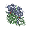
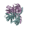
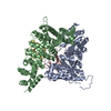
- PDB-7q84: Crystal structure of human peroxisomal acyl-Co-A oxidase 1a, apo-form -
+ Open data

- Basic information
| Entry | Database: PDB / ID: 7q84 | |||||||||
|---|---|---|---|---|---|---|---|---|---|---|
| Title | Crystal structure of human peroxisomal acyl-Co-A oxidase 1a, apo-form | |||||||||
 Components Components | Isoform 2 of Peroxisomal acyl-coenzyme A oxidase 1 | |||||||||
 Keywords Keywords |  OXIDOREDUCTASE / acyl-Co-A oxidase / peroxisomal enzyme OXIDOREDUCTASE / acyl-Co-A oxidase / peroxisomal enzyme | |||||||||
| Function / homology | DI(HYDROXYETHYL)ETHER / TRIETHYLENE GLYCOL Function and homology information Function and homology information | |||||||||
| Biological species |  Homo sapiens (human) Homo sapiens (human) | |||||||||
| Method | X-RAY DIFFRACTION / SYNCHROTRON / MOLECULAR REPLACEMENT / Resolution: 2 Å | |||||||||
 Authors Authors | Sonani, R.R. / Blat, A. / Dubin, G. | |||||||||
| Funding support | 2items
| |||||||||
 Citation Citation | Journal: Int.J.Biol.Macromol. / Year: 2022 Title: Crystal structures of apo- and FAD-bound human peroxisomal acyl-CoA oxidase provide mechanistic basis explaining clinical observations. Authors: Sonani, R.R. / Blat, A. / Dubin, G. | |||||||||
| History |
|
- Structure visualization
| Structure viewer | Molecule: MolmilJmol/JSmol |
|---|
- Downloads & links
-Download
| PDBx/mmCIF format | 7q84.cif.gz | 505.7 KB | Display | PDBx/mmCIF format |
|---|---|---|---|---|
| PDB format | pdb7q84.ent.gz | 413.9 KB | Display | PDB format |
| PDBx/mmJSON format | 7q84.json.gz | Tree view | PDBx/mmJSON format | |
| Others |  Other downloads Other downloads |
-Validation report
| Arichive directory | https://data.pdbj.org/pub/pdb/validation_reports/q8/7q84ftp://data.pdbj.org/pub/pdb/validation_reports/q8/7q84 | HTTPS FTP |
|---|
-Related structure data
| Related structure data | 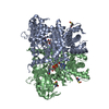 7q86C 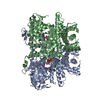 1is2S S: Starting model for refinement C: citing same article ( |
|---|---|
| Similar structure data |


-Links
 PDBj
PDBj- Assembly
| Deposited unit | 
| ||||||||
|---|---|---|---|---|---|---|---|---|---|
| 1 |
| ||||||||
| Unit cell |
|
-Components
-Protein , 1 types, 2 molecules AB
| #1: Protein | Mass: 75753.641 Da / Num. of mol.: 2 Source method: isolated from a genetically manipulated source Source: (gene. exp.) Homo sapiens (human) / Gene: ACOX1, ACOX / Production host:  Escherichia coli (E. coli) / References: acyl-CoA oxidase Escherichia coli (E. coli) / References: acyl-CoA oxidase |
|---|
-Non-polymers , 5 types, 508 molecules 


| #2: Chemical | Polyethylene glycol Mass: 150.173 Da / Num. of mol.: 2 / Source method: obtained synthetically / Formula: C6H14O4 Mass: 150.173 Da / Num. of mol.: 2 / Source method: obtained synthetically / Formula: C6H14O4#3: Chemical | ChemComp-EDO / Ethylene glycol Mass: 62.068 Da / Num. of mol.: 10 / Source method: obtained synthetically / Formula: C2H6O2 Mass: 62.068 Da / Num. of mol.: 10 / Source method: obtained synthetically / Formula: C2H6O2#4: Chemical | ChemComp-PEG / | Diethylene glycol Mass: 106.120 Da / Num. of mol.: 1 / Source method: obtained synthetically / Formula: C4H10O3 Mass: 106.120 Da / Num. of mol.: 1 / Source method: obtained synthetically / Formula: C4H10O3#5: Chemical | Glycerol Mass: 92.094 Da / Num. of mol.: 3 / Source method: obtained synthetically / Formula: C3H8O3 Mass: 92.094 Da / Num. of mol.: 3 / Source method: obtained synthetically / Formula: C3H8O3#6: Water | ChemComp-HOH / | WaterMass: 18.015 Da / Num. of mol.: 492 / Source method: isolated from a natural source / Formula: H2O |
|---|
-Details
| Has ligand of interest | N |
|---|
-Experimental details
-Experiment
| Experiment | Method: X-RAY DIFFRACTION / Number of used crystals: 1 |
|---|
- Sample preparation
| Crystal | Density Matthews: 2.73 Å3/Da / Density % sol: 54.96 % |
|---|---|
| Crystal grow | Temperature: 298.15 K / Method: vapor diffusion, sitting drop Details: 12.5 % PEG 8000, 0.09 M ammonium sulfate, 0.05 M sodium cacodylate (pH 6.0) |
-Data collection
| Diffraction | Mean temperature: 80 K / Serial crystal experiment: N |
|---|---|
| Diffraction source | Source: SYNCHROTRON / Site: PETRA III, EMBL c/o DESY  / Beamline: P13 (MX1) / Wavelength: 1.0332 Å / Beamline: P13 (MX1) / Wavelength: 1.0332 Å |
| Detector | Type: DECTRIS EIGER X 4M / Detector: PIXEL / Date: May 1, 2021 |
| Radiation | Protocol: SINGLE WAVELENGTH / Monochromatic (M) / Laue (L): M / Scattering type: x-ray |
| Radiation wavelength | Wavelength: 1.0332 Å / Relative weight: 1 |
| Reflection | Resolution: 2→48.18 Å / Num. obs: 110023 / % possible obs: 99.03 % / Redundancy: 13.9 % / CC1/2: 0.999 / CC star: 1 / Rmerge(I) obs: 0.092 / Rpim(I) all: 0.026 / Rrim(I) all: 0.096 / Net I/σ(I): 17.38 |
| Reflection shell | Resolution: 2→2.071 Å / Rmerge(I) obs: 1.319 / Num. unique obs: 10875 / CC1/2: 0.86 / CC star: 0.962 / Rpim(I) all: 0.358 / Rrim(I) all: 1.367 / % possible all: 99.35 |
- Processing
| Software |
| |||||||||||||||||||||||||||||||||||||||||||||||||||||||||||||||||||||||||||
|---|---|---|---|---|---|---|---|---|---|---|---|---|---|---|---|---|---|---|---|---|---|---|---|---|---|---|---|---|---|---|---|---|---|---|---|---|---|---|---|---|---|---|---|---|---|---|---|---|---|---|---|---|---|---|---|---|---|---|---|---|---|---|---|---|---|---|---|---|---|---|---|---|---|---|---|---|
| Refinement | Method to determine structure: MOLECULAR REPLACEMENT Starting model: 1is2 Resolution: 2→48.23 Å / Cor.coef. Fo:Fc: 0.968 / Cor.coef. Fo:Fc free: 0.955 / SU B: 10.326 / SU ML: 0.133 / Cross valid method: THROUGHOUT / σ(F): 0 / ESU R: 0.154 / ESU R Free: 0.143 / Stereochemistry target values: MAXIMUM LIKELIHOOD Details: HYDROGENS HAVE BEEN ADDED IN THE RIDING POSITIONS U VALUES : WITH TLS ADDED
| |||||||||||||||||||||||||||||||||||||||||||||||||||||||||||||||||||||||||||
| Solvent computation | Ion probe radii: 0.8 Å / Shrinkage radii: 0.8 Å / VDW probe radii: 1.2 Å / Solvent model: MASK | |||||||||||||||||||||||||||||||||||||||||||||||||||||||||||||||||||||||||||
| Displacement parameters | Biso max: 153.54 Å2 / Biso mean: 47.835 Å2 / Biso min: 20 Å2
| |||||||||||||||||||||||||||||||||||||||||||||||||||||||||||||||||||||||||||
| Refinement step | Cycle: final / Resolution: 2→48.23 Å
| |||||||||||||||||||||||||||||||||||||||||||||||||||||||||||||||||||||||||||
| Refine LS restraints |
| |||||||||||||||||||||||||||||||||||||||||||||||||||||||||||||||||||||||||||
| LS refinement shell | Resolution: 2→2.052 Å / Rfactor Rfree error: 0 / Total num. of bins used: 20
| |||||||||||||||||||||||||||||||||||||||||||||||||||||||||||||||||||||||||||
| Refinement TLS params. | Method: refined / Refine-ID: X-RAY DIFFRACTION
| |||||||||||||||||||||||||||||||||||||||||||||||||||||||||||||||||||||||||||
| Refinement TLS group |
|
