 Movie
Movie Controller

[English] 日本語
 Yorodumi
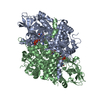
- PDB-1is2: Crystal Structure of Peroxisomal Acyl-CoA Oxidase-II from Rat Liver -
+ Open data

- Basic information
| Entry | Database: PDB / ID: 1is2 | ||||||
|---|---|---|---|---|---|---|---|
| Title | Crystal Structure of Peroxisomal Acyl-CoA Oxidase-II from Rat Liver | ||||||
 Components Components | acyl-CoA oxidase | ||||||
 Keywords Keywords | OXIDOREDUCTASE / FAD | ||||||
| Function / homology |  Function and homology information Function and homology informationvery long-chain fatty acid beta-oxidation / alpha-linolenic acid (ALA) metabolism / palmitoyl-CoA oxidase activity / acyl-CoA oxidase / Beta-oxidation of very long chain fatty acids / acyl-CoA oxidase activity / fatty acid beta-oxidation using acyl-CoA oxidase / Peroxisomal protein import / very long-chain fatty acid metabolic process / fatty acid catabolic process ...very long-chain fatty acid beta-oxidation / alpha-linolenic acid (ALA) metabolism / palmitoyl-CoA oxidase activity / acyl-CoA oxidase / Beta-oxidation of very long chain fatty acids / acyl-CoA oxidase activity / fatty acid beta-oxidation using acyl-CoA oxidase / Peroxisomal protein import / very long-chain fatty acid metabolic process / fatty acid catabolic process / hydrogen peroxide biosynthetic process / peroxisomal membrane / prostaglandin metabolic process / lipid homeostasis / fatty acid oxidation / peroxisomal matrix / FAD binding / generation of precursor metabolites and energy / fatty acid binding / PDZ domain binding / lipid metabolic process / peroxisome / flavin adenine dinucleotide binding / spermatogenesis / protein homodimerization activity / cytoplasmSimilarity search - Function | ||||||
| Biological species |  Rattus norvegicus (Norway rat) Rattus norvegicus (Norway rat) | ||||||
| Method | X-RAY DIFFRACTION / SYNCHROTRON / MIRAS / Resolution: 2.2 Å | ||||||
 Authors Authors | Nakajima, Y. / Miyahara, I. / Hirotsu, K. | ||||||
 Citation Citation | Journal: J.Biochem. / Year: 2002 Title: Three-dimensional structure of the flavoenzyme acyl-CoA oxidase-II from rat liver, the peroxisomal counterpart of mitochondrial acyl-CoA dehydrogenase. Authors: Nakajima, Y. / Miyahara, I. / Hirotsu, K. / Nishina, Y. / Shiga, K. / Setoyama, C. / Tamaoki, H. / Miura, R. | ||||||
| History |
|
- Structure visualization
| Structure viewer | Molecule: MolmilJmol/JSmol |
|---|
- Downloads & links
-Download
| PDBx/mmCIF format | 1is2.cif.gz | 264.9 KB | Display | PDBx/mmCIF format |
|---|---|---|---|---|
| PDB format | pdb1is2.ent.gz | 211.9 KB | Display | PDB format |
| PDBx/mmJSON format | 1is2.json.gz | Tree view | PDBx/mmJSON format | |
| Others |  Other downloads Other downloads |
-Validation report
| Arichive directory | https://data.pdbj.org/pub/pdb/validation_reports/is/1is2ftp://data.pdbj.org/pub/pdb/validation_reports/is/1is2 | HTTPS FTP |
|---|
-Related structure data
| Similar structure data |
|---|


-Links
 PDBj
PDBj- Assembly
| Deposited unit | 
| ||||||||
|---|---|---|---|---|---|---|---|---|---|
| 1 |
| ||||||||
| Unit cell |
| ||||||||
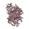
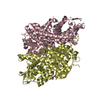
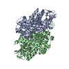
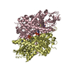
| Details | The biological assembly is a dimer in the asymmetric unit. |
-Components
| #1: Protein | Mass: 74784.531 Da / Num. of mol.: 2 Source method: isolated from a genetically manipulated source Source: (gene. exp.) Rattus norvegicus (Norway rat) / Plasmid: pET11d / Production host:  Escherichia coli (E. coli) / References: UniProt: P07872, acyl-CoA oxidase Escherichia coli (E. coli) / References: UniProt: P07872, acyl-CoA oxidase#2: Chemical | Flavin adenine dinucleotide  Mass: 785.550 Da / Num. of mol.: 2 / Source method: obtained synthetically / Formula: C27H33N9O15P2 / Comment: FAD*YM Mass: 785.550 Da / Num. of mol.: 2 / Source method: obtained synthetically / Formula: C27H33N9O15P2 / Comment: FAD*YM#3: Water | ChemComp-HOH / | Water Mass: 18.015 Da / Num. of mol.: 443 / Source method: isolated from a natural source / Formula: H2O Mass: 18.015 Da / Num. of mol.: 443 / Source method: isolated from a natural source / Formula: H2O |
|---|
-Experimental details
-Experiment
| Experiment | Method: X-RAY DIFFRACTION / Number of used crystals: 1 |
|---|
- Sample preparation
| Crystal | Density Matthews: 2.36 Å3/Da / Density % sol: 47.92 % | ||||||||||||||||||||||||||||||||||||
|---|---|---|---|---|---|---|---|---|---|---|---|---|---|---|---|---|---|---|---|---|---|---|---|---|---|---|---|---|---|---|---|---|---|---|---|---|---|
| Crystal grow | Temperature: 293 K / Method: vapor diffusion, hanging drop / pH: 7.4 Details: PEG 20000, potassium phosphate, pH 7.4, VAPOR DIFFUSION, HANGING DROP, temperature 293K | ||||||||||||||||||||||||||||||||||||
| Crystal grow | *PLUS | ||||||||||||||||||||||||||||||||||||
| Components of the solutions | *PLUS
|
-Data collection
| Diffraction | Mean temperature: 100 K |
|---|---|
| Diffraction source | Source: SYNCHROTRON / Site: SPring-8  / Beamline: BL41XU / Wavelength: 1 Å / Beamline: BL41XU / Wavelength: 1 Å |
| Detector | Type: MAR CCD 165 mm / Detector: CCD / Date: Jun 8, 2001 |
| Radiation | Protocol: SINGLE WAVELENGTH / Monochromatic (M) / Laue (L): M / Scattering type: x-ray |
| Radiation wavelength | Wavelength: 1 Å / Relative weight: 1 |
| Reflection | Resolution: 2.2→20 Å / Num. all: 69647 / Num. obs: 69647 / % possible obs: 94.7 % / Observed criterion σ(F): 0 / Observed criterion σ(I): 0 / Rmerge(I) obs: 0.06 / Net I/σ(I): 16 |
| Reflection shell | Resolution: 2.2→2.27 Å / Rmerge(I) obs: 0.283 / Mean I/σ(I) obs: 2.1 / Num. unique all: 6152 / % possible all: 85.3 |
| Reflection | *PLUS Highest resolution: 2.2 Å / Num. measured all: 344046 / Rmerge(I) obs: 0.06 |
| Reflection shell | *PLUS % possible obs: 85.3 % / Num. unique obs: 6152 / Num. measured obs: 22267 / Rmerge(I) obs: 0.283 |
- Processing
| Software |
| |||||||||||||||||||||||||
|---|---|---|---|---|---|---|---|---|---|---|---|---|---|---|---|---|---|---|---|---|---|---|---|---|---|---|
| Refinement | Method to determine structure: MIRAS / Resolution: 2.2→10 Å / σ(F): 2 / Stereochemistry target values: RANDOM
| |||||||||||||||||||||||||
| Displacement parameters | Biso mean: 32.8 Å2 | |||||||||||||||||||||||||
| Refine analyze | Luzzati coordinate error obs: 0.28 Å / Luzzati d res low obs: 5 Å / Luzzati sigma a obs: 0.21 Å | |||||||||||||||||||||||||
| Refinement step | Cycle: LAST / Resolution: 2.2→10 Å
| |||||||||||||||||||||||||
| Refine LS restraints |
| |||||||||||||||||||||||||
| Refinement | *PLUS Rfactor obs: 0.209 / Rfactor Rfree: 0.26 / Rfactor Rwork: 0.209 | |||||||||||||||||||||||||
| Solvent computation | *PLUS | |||||||||||||||||||||||||
| Displacement parameters | *PLUS | |||||||||||||||||||||||||
| Refine LS restraints | *PLUS
|
