 Movie
Movie Controller

[English] 日本語
 Yorodumi
- PDB-7q7c: Room temperature structure of the human Serine/Threonine Kinase 1... -
+ Open data

- Basic information
| Entry | Database: PDB / ID: 7q7c | ||||||
|---|---|---|---|---|---|---|---|
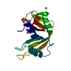
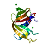
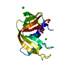
| Title | Room temperature structure of the human Serine/Threonine Kinase 17B (STK17B/DRAK2) in complex with ADP at atmospheric pressure | ||||||
 Components Components | Serine/threonine-protein kinase 17B | ||||||
 Keywords Keywords |  TRANSFERASE / HPMX / high-pressure macromolecular crystallography / sapphire capillary / room temperature TRANSFERASE / HPMX / high-pressure macromolecular crystallography / sapphire capillary / room temperature | ||||||
| Function / homology |  Function and homology information Function and homology informationpositive regulation of fibroblast apoptotic process / Flemming body / endoplasmic reticulum-Golgi intermediate compartment / actin cytoskeleton / protein autophosphorylation / non-specific serine/threonine protein kinase / protein kinase activity / intracellular signal transduction / positive regulation of apoptotic process / protein phosphorylation ...positive regulation of fibroblast apoptotic process / Flemming body / endoplasmic reticulum-Golgi intermediate compartment / actin cytoskeleton / protein autophosphorylation / non-specific serine/threonine protein kinase / protein kinase activity / intracellular signal transduction / positive regulation of apoptotic process / protein phosphorylation / protein serine kinase activity / protein serine/threonine kinase activity / apoptotic process / nucleoplasm / ATP binding / nucleus / plasma membraneSimilarity search - Function | ||||||
| Biological species |  Homo sapiens (human) Homo sapiens (human) | ||||||
| Method | X-RAY DIFFRACTION / SYNCHROTRON / MOLECULAR REPLACEMENT / Resolution: 2.85 Å | ||||||
 Authors Authors | Lieske, J. / Guenther, S. / Saouane, S. / Meents, A. | ||||||
| Funding support |  Germany, 1items Germany, 1items
| ||||||
 Citation Citation | Journal: To Be Published Title: Room temperature structure of the human Serine/Threonine Kinase 17B (STK17B/DRAK2) in complex with ADP at atmospheric pressure Authors: Lieske, J. / Saouane, S. / Guenther, S. / Meyer, J. / Pakendorf, T. / Reime, B. / Burkhardt, A. / Crosas, E. / Hakanpaeae, J. / Stachnik, K. / Sieg, J. / Rarey, M. / Abdellatif, M.H. / ...Authors: Lieske, J. / Saouane, S. / Guenther, S. / Meyer, J. / Pakendorf, T. / Reime, B. / Burkhardt, A. / Crosas, E. / Hakanpaeae, J. / Stachnik, K. / Sieg, J. / Rarey, M. / Abdellatif, M.H. / Gabdulkhakov, A.G. / Selikhanov, G.K. / Chapman, H.N. / Meents, A. | ||||||
| History |
|
- Structure visualization
| Structure viewer | Molecule: MolmilJmol/JSmol |
|---|
- Downloads & links
-Download
| PDBx/mmCIF format | 7q7c.cif.gz | 210.4 KB | Display | PDBx/mmCIF format |
|---|---|---|---|---|
| PDB format | pdb7q7c.ent.gz | 141.5 KB | Display | PDB format |
| PDBx/mmJSON format | 7q7c.json.gz | Tree view | PDBx/mmJSON format | |
| Others |  Other downloads Other downloads |
-Validation report
| Arichive directory | https://data.pdbj.org/pub/pdb/validation_reports/q7/7q7cftp://data.pdbj.org/pub/pdb/validation_reports/q7/7q7c | HTTPS FTP |
|---|
-Related structure data
| Related structure data |  6qf4S S: Starting model for refinement |
|---|---|
| Similar structure data |


-Links
 PDBj
PDBj- Assembly
| Deposited unit | 
| ||||||||||||
|---|---|---|---|---|---|---|---|---|---|---|---|---|---|
| 1 |
| ||||||||||||
| Unit cell |
|
-Components
| #1: Protein | Mass: 37327.086 Da / Num. of mol.: 1 Source method: isolated from a genetically manipulated source Source: (gene. exp.) Homo sapiens (human) / Gene: STK17B, DRAK2 / Production host:  Escherichia coli (E. coli) Escherichia coli (E. coli)References: UniProt: O94768, non-specific serine/threonine protein kinase |
|---|---|
| #2: Chemical | ChemComp-MG /   Mass: 24.305 Da / Num. of mol.: 1 / Source method: obtained synthetically / Formula: Mg / Feature type: SUBJECT OF INVESTIGATION Mass: 24.305 Da / Num. of mol.: 1 / Source method: obtained synthetically / Formula: Mg / Feature type: SUBJECT OF INVESTIGATION |
| #3: Chemical | ChemComp-ADP / Adenosine diphosphate  Mass: 427.201 Da / Num. of mol.: 1 / Source method: obtained synthetically / Formula: C10H15N5O10P2 / Feature type: SUBJECT OF INVESTIGATION / Comment: ADP, energy-carrying molecule*YM Mass: 427.201 Da / Num. of mol.: 1 / Source method: obtained synthetically / Formula: C10H15N5O10P2 / Feature type: SUBJECT OF INVESTIGATION / Comment: ADP, energy-carrying molecule*YM |
| #4: Water | ChemComp-HOH / Water Mass: 18.015 Da / Num. of mol.: 6 / Source method: isolated from a natural source / Formula: H2O Mass: 18.015 Da / Num. of mol.: 6 / Source method: isolated from a natural source / Formula: H2O |
| Has ligand of interest | Y |
-Experimental details
-Experiment
| Experiment | Method: X-RAY DIFFRACTION / Number of used crystals: 1 |
|---|
- Sample preparation
| Crystal | Density Matthews: 2.78 Å3/Da / Density % sol: 55.82 % |
|---|---|
| Crystal grow | Temperature: 295 K / Method: batch mode Details: 20 mg/ml protein in 25 mM HEPES pH 7.5, 500 mM NaCl, 0.5 mM TCEP was mixed 1:1.5 with precipitant solution (0.1 M HEPES 6.5, 0.2M ammonium acetate, 20% PEG 3350, 50mM sodium/potassium ...Details: 20 mg/ml protein in 25 mM HEPES pH 7.5, 500 mM NaCl, 0.5 mM TCEP was mixed 1:1.5 with precipitant solution (0.1 M HEPES 6.5, 0.2M ammonium acetate, 20% PEG 3350, 50mM sodium/potassium tartrate, 0.8mM quercetin). Batch grown crystals were subsequently mixed 1:1 with 100 mM ATP/50mM magnesium chloride in DRAK2 crystallization buffer and incubated for half an hour. PH range: 6.5-7.5 |
-Data collection
| Diffraction | Mean temperature: 295 K / Serial crystal experiment: N |
|---|---|
| Diffraction source | Source: SYNCHROTRON / Site: PETRA III, DESY / Beamline: P11 / Wavelength: 0.979 Å |
| Detector | Type: DECTRIS PILATUS3 X CdTe 2M / Detector: PIXEL / Date: Nov 15, 2019 |
| Diffraction measurement | Specimen support: sapphire capillary |
| Radiation | Protocol: SINGLE WAVELENGTH / Monochromatic (M) / Laue (L): M / Scattering type: x-ray |
| Radiation wavelength | Wavelength: 0.979 Å / Relative weight: 1 |
| Reflection | Resolution: 2.85→47.98 Å / Num. obs: 10282 / % possible obs: 98.91 % / Redundancy: 7.8 % / Biso Wilson estimate: 92.37 Å2 / CC1/2: 0.991 / CC star: 0.998 / Rmerge(I) obs: 0.201 / Rpim(I) all: 0.074 / Rrim(I) all: 0.215 / Net I/σ(I): 5.93 |
| Reflection shell | Resolution: 2.85→2.952 Å / Redundancy: 5.5 % / Rmerge(I) obs: 1.085 / Mean I/σ(I) obs: 0.99 / Num. unique obs: 982 / CC1/2: 0.457 / CC star: 0.792 / Rpim(I) all: 0.486 / Rrim(I) all: 1.195 / % possible all: 97.31 |
| Cell measurement | Pressure: 101.325 kPa |
- Processing
| Software |
| ||||||||||||||||||||||||||||||||||||||||||||||||||||||||||||||||||||||||||||||||||||||||||||||||||||
|---|---|---|---|---|---|---|---|---|---|---|---|---|---|---|---|---|---|---|---|---|---|---|---|---|---|---|---|---|---|---|---|---|---|---|---|---|---|---|---|---|---|---|---|---|---|---|---|---|---|---|---|---|---|---|---|---|---|---|---|---|---|---|---|---|---|---|---|---|---|---|---|---|---|---|---|---|---|---|---|---|---|---|---|---|---|---|---|---|---|---|---|---|---|---|---|---|---|---|---|---|---|
| Refinement | Method to determine structure: MOLECULAR REPLACEMENT Starting model: 6QF4 Resolution: 2.85→47.98 Å / SU ML: 0.4636 / Cross valid method: FREE R-VALUE / σ(F): 1.33 / Phase error: 25.9839 / Stereochemistry target values: GeoStd + Monomer Library
| ||||||||||||||||||||||||||||||||||||||||||||||||||||||||||||||||||||||||||||||||||||||||||||||||||||
| Solvent computation | Shrinkage radii: 0.9 Å / VDW probe radii: 1.11 Å / Solvent model: FLAT BULK SOLVENT MODEL | ||||||||||||||||||||||||||||||||||||||||||||||||||||||||||||||||||||||||||||||||||||||||||||||||||||
| Displacement parameters | Biso mean: 97.15 Å2 | ||||||||||||||||||||||||||||||||||||||||||||||||||||||||||||||||||||||||||||||||||||||||||||||||||||
| Refinement step | Cycle: LAST / Resolution: 2.85→47.98 Å
| ||||||||||||||||||||||||||||||||||||||||||||||||||||||||||||||||||||||||||||||||||||||||||||||||||||
| Refine LS restraints |
| ||||||||||||||||||||||||||||||||||||||||||||||||||||||||||||||||||||||||||||||||||||||||||||||||||||
| LS refinement shell |
| ||||||||||||||||||||||||||||||||||||||||||||||||||||||||||||||||||||||||||||||||||||||||||||||||||||
| Refinement TLS params. | Method: refined / Refine-ID: X-RAY DIFFRACTION
| ||||||||||||||||||||||||||||||||||||||||||||||||||||||||||||||||||||||||||||||||||||||||||||||||||||
| Refinement TLS group |
|
