 Movie
Movie Controller


+ Open data

- Basic information
| Entry | Database: PDB / ID: 7py2 | ||||||
|---|---|---|---|---|---|---|---|
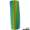
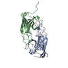
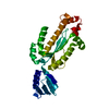
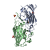
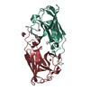
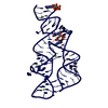
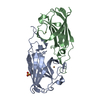
| Title | Structure of pathological TDP-43 filaments from ALS with FTLD | ||||||
 Components Components | TAR DNA-binding protein 43 | ||||||
 Keywords Keywords | PROTEIN FIBRIL / TDP-43 / ALS / amyotrophic lateral sclerosis / FTD / frontotemporal dementia / FTLD / frontotemporal lobar degeneration / amyloid / filament / fibril / neurodegenerative / neurodegeneration | ||||||
| Function / homology |  Function and homology information Function and homology informationnuclear inner membrane organization / interchromatin granule / perichromatin fibrils / 3'-UTR-mediated mRNA destabilization / 3'-UTR-mediated mRNA stabilization / intracellular non-membrane-bounded organelle / negative regulation by host of viral transcription / pre-mRNA intronic binding / response to endoplasmic reticulum stress / RNA splicing ...nuclear inner membrane organization / interchromatin granule / perichromatin fibrils / 3'-UTR-mediated mRNA destabilization / 3'-UTR-mediated mRNA stabilization / intracellular non-membrane-bounded organelle / negative regulation by host of viral transcription / pre-mRNA intronic binding / response to endoplasmic reticulum stress / RNA splicing / negative regulation of protein phosphorylation / molecular condensate scaffold activity / mRNA 3'-UTR binding / regulation of protein stability / regulation of circadian rhythm / positive regulation of insulin secretion / mRNA processing / cytoplasmic stress granule / positive regulation of protein import into nucleus / rhythmic process / regulation of gene expression / double-stranded DNA binding / regulation of apoptotic process / amyloid fibril formation / regulation of cell cycle / nuclear speck / RNA polymerase II cis-regulatory region sequence-specific DNA binding / negative regulation of gene expression / lipid binding / mitochondrion / DNA binding / RNA binding / nucleoplasm / identical protein binding / nucleusSimilarity search - Function | ||||||
| Biological species |  Homo sapiens (human) Homo sapiens (human) | ||||||
| Method | ELECTRON MICROSCOPY / helical reconstruction / cryo EM / Resolution: 2.59 Å | ||||||
 Authors Authors | Arseni, D. / Hasegawa, H. / Murzin, A.G. / Kametani, F. / Arai, M. / Yoshida, M. / Falcon, B. | ||||||
| Funding support |  United Kingdom, 1items United Kingdom, 1items
| ||||||
 Citation Citation | Journal: Nature / Year: 2022 Title: Structure of pathological TDP-43 filaments from ALS with FTLD. Authors: Diana Arseni / Masato Hasegawa / Alexey G Murzin / Fuyuki Kametani / Makoto Arai / Mari Yoshida / Benjamin Ryskeldi-Falcon /  Abstract: The abnormal aggregation of TAR DNA-binding protein 43 kDa (TDP-43) in neurons and glia is the defining pathological hallmark of the neurodegenerative disease amyotrophic lateral sclerosis (ALS) ...The abnormal aggregation of TAR DNA-binding protein 43 kDa (TDP-43) in neurons and glia is the defining pathological hallmark of the neurodegenerative disease amyotrophic lateral sclerosis (ALS) and multiple forms of frontotemporal lobar degeneration (FTLD). It is also common in other diseases, including Alzheimer's and Parkinson's. No disease-modifying therapies exist for these conditions and early diagnosis is not possible. The structures of pathological TDP-43 aggregates are unknown. Here we used cryo-electron microscopy to determine the structures of aggregated TDP-43 in the frontal and motor cortices of an individual who had ALS with FTLD and from the frontal cortex of a second individual with the same diagnosis. An identical amyloid-like filament structure comprising a single protofilament was found in both brain regions and individuals. The ordered filament core spans residues 282-360 in the TDP-43 low-complexity domain and adopts a previously undescribed double-spiral-shaped fold, which shows no similarity to those of TDP-43 filaments formed in vitro. An abundance of glycine and neutral polar residues facilitates numerous turns and restricts β-strand length, which results in an absence of β-sheet stacking that is associated with cross-β amyloid structure. An uneven distribution of residues gives rise to structurally and chemically distinct surfaces that face external densities and suggest possible ligand-binding sites. This work enhances our understanding of the molecular pathogenesis of ALS and FTLD and informs the development of diagnostic and therapeutic agents that target aggregated TDP-43. | ||||||
| History |
|
- Structure visualization
| Movie |
Movie viewer |
|---|---|
| Structure viewer | Molecule: MolmilJmol/JSmol |
- Downloads & links
-Download
| PDBx/mmCIF format | 7py2.cif.gz | 80.5 KB | Display | PDBx/mmCIF format |
|---|---|---|---|---|
| PDB format | pdb7py2.ent.gz | 54.3 KB | Display | PDB format |
| PDBx/mmJSON format | 7py2.json.gz | Tree view | PDBx/mmJSON format | |
| Others |  Other downloads Other downloads |
-Validation report
| Arichive directory | https://data.pdbj.org/pub/pdb/validation_reports/py/7py2ftp://data.pdbj.org/pub/pdb/validation_reports/py/7py2 | HTTPS FTP |
|---|
-Related structure data
| Related structure data | 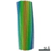 13708MC M: map data used to model this data C: citing same article ( |
|---|---|
| Similar structure data | |
| EM raw data | EMPIAR-10830 (Title: Structure of pathological TDP-43 filaments from ALS with FTLD (Individual 1, frontal cortex) Data size: 3.9 TB Data #1: Unaligned multiframe movies [micrographs - multiframe]) |


-Links
 PDBj
PDBj
- Assembly
| Deposited unit | 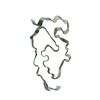
| ||||||||||||||||||||
|---|---|---|---|---|---|---|---|---|---|---|---|---|---|---|---|---|---|---|---|---|---|
| 1 |
| ||||||||||||||||||||
| Noncrystallographic symmetry (NCS) | NCS oper:
|
-Components
| #1: Protein | / TDP-43 Mass: 44784.742 Da / Num. of mol.: 4 / Source method: isolated from a natural source / Source: (natural) Homo sapiens (human) / Tissue: Brain / References: UniProt: Q13148 |
|---|
-Experimental details
-Experiment
| Experiment | Method: ELECTRON MICROSCOPY |
|---|---|
| EM experiment | Aggregation state: FILAMENT / 3D reconstruction method: helical reconstruction |
- Sample preparation
| Component | Name: Pathological TDP-43 filaments extracted from the frontal cortex of an individual that succumbed to ALS with FTLD. Type: TISSUE / Entity ID: all / Source: NATURAL |
|---|---|
| Source (natural) | Organism: Homo sapiens (human) / Cellular location: Cytoplasm / Tissue: Brain |
| Buffer solution | pH: 7.4 |
| Specimen | Embedding applied: NO / Shadowing applied: NO / Staining applied: NO / Vitrification applied: YES |
| Vitrification | Cryogen name: ETHANE |
- Electron microscopy imaging
| Experimental equipment |  Model: Titan Krios / Image courtesy: FEI Company |
|---|---|
| Microscopy | Model: FEI TITAN KRIOS |
| Electron gun | Electron source: FIELD EMISSION GUN / Accelerating voltage: 300 kV / Illumination mode: FLOOD BEAM |
| Electron lens | Mode: BRIGHT FIELDBright-field microscopy |
| Image recording | Electron dose: 39.7 e/Å2 / Film or detector model: GATAN K3 (6k x 4k) |
- Processing
| Software | Name: REFMAC / Version: 5.8.0272 / Classification: refinement | ||||||||||||||||||||||||||||||||||||||||||||||||||||||||||||||||||||||||||||||||||||||||||||||||||||||||||
|---|---|---|---|---|---|---|---|---|---|---|---|---|---|---|---|---|---|---|---|---|---|---|---|---|---|---|---|---|---|---|---|---|---|---|---|---|---|---|---|---|---|---|---|---|---|---|---|---|---|---|---|---|---|---|---|---|---|---|---|---|---|---|---|---|---|---|---|---|---|---|---|---|---|---|---|---|---|---|---|---|---|---|---|---|---|---|---|---|---|---|---|---|---|---|---|---|---|---|---|---|---|---|---|---|---|---|---|
| CTF correction | Type: PHASE FLIPPING AND AMPLITUDE CORRECTION | ||||||||||||||||||||||||||||||||||||||||||||||||||||||||||||||||||||||||||||||||||||||||||||||||||||||||||
| Helical symmerty | Angular rotation/subunit: 1.422 ° / Axial rise/subunit: 4.842 Å / Axial symmetry: C1 | ||||||||||||||||||||||||||||||||||||||||||||||||||||||||||||||||||||||||||||||||||||||||||||||||||||||||||
| 3D reconstruction | Resolution: 2.59 Å / Resolution method: FSC 0.143 CUT-OFF / Num. of particles: 308822 / Symmetry type: HELICAL | ||||||||||||||||||||||||||||||||||||||||||||||||||||||||||||||||||||||||||||||||||||||||||||||||||||||||||
| Refinement | Resolution: 2.6→85.56 Å / Cor.coef. Fo:Fc: 0.721 / SU B: 7.738 / SU ML: 0.161 / ESU R: 0.151 Stereochemistry target values: MAXIMUM LIKELIHOOD WITH PHASES Details: HYDROGENS HAVE BEEN ADDED IN THE RIDING POSITIONS | ||||||||||||||||||||||||||||||||||||||||||||||||||||||||||||||||||||||||||||||||||||||||||||||||||||||||||
| Solvent computation | Solvent model: PARAMETERS FOR MASK CACLULATION | ||||||||||||||||||||||||||||||||||||||||||||||||||||||||||||||||||||||||||||||||||||||||||||||||||||||||||
| Displacement parameters | Biso mean: 64.971 Å2 | ||||||||||||||||||||||||||||||||||||||||||||||||||||||||||||||||||||||||||||||||||||||||||||||||||||||||||
| Refinement step | Cycle: 1 / Total: 538 | ||||||||||||||||||||||||||||||||||||||||||||||||||||||||||||||||||||||||||||||||||||||||||||||||||||||||||
| Refine LS restraints |
|