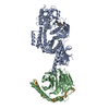
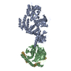
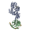
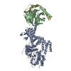
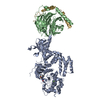
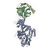
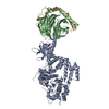
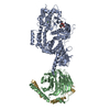
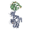
Entry Database : PDB / ID : 7pwdTitle Structure of an inhibited GRK2-G-beta and G-gamma complex (Guanine nucleotide-binding protein ...) x 2 Beta-adrenergic receptor kinase 1 Keywords / / / / / Function / homology Function Domain/homology Component
/ / / / / / / / / / / / / / / / / / / / / / / / / / / / / / / / / / / / / / / / / / / / / / / / / / / / / / / / / / / / / / / / / / / / / / / / / / / / / / / / / / / / / / / / / / / / / / / / / / / / / / / / / / / / / / / / / / / / / / / / / / / / / / / / / / / / / / Biological species Homo sapiens (human)Bos taurus (cattle)Method / / / / Resolution : 2.6 Å Authors Faucher, N. / Tauchert, M.J. / Konz Makino, D.L. / Vuillard, L.M. Funding support 1items Organization Grant number Country Not funded
Journal : Monatsh Chem / Year : 2022Title : Synthesis of thieno[2,3-c]pyridine derived GRK2 inhibitorsAuthors : Balo, T. / Sapi, A. / Kiss, A. / Raimbaud, E. / Paysant, J. / Cattin, M.E. / Berger, S. / Kotschy, A. / Faucher, N. History Deposition Oct 6, 2021 Deposition site / Processing site Revision 1.0 Oct 20, 2021 Provider / Type Revision 1.1 May 18, 2022 Group / Refinement description / Category / citation_author / refineItem _citation.journal_abbrev / _citation.journal_id_CSD ... _citation.journal_abbrev / _citation.journal_id_CSD / _citation.pdbx_database_id_DOI / _citation.title / _citation.year / _refine.pdbx_diffrn_id
Show all Show less
 Movie
Movie Controller


 Open data
 Basic information
 Components
 Keywords
 TRANSFERASE / Proteros /
  Function and homology information

 Authors
 Citation
 Structure visualization
 Downloads & links
 Other downloads
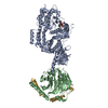
 PDBj
PDBj


 Assembly


 Mass: 351.250 Da / Num. of mol.: 1 / Source method: obtained synthetically / Formula: C16H12Cl2N2OS / Feature type: SUBJECT OF INVESTIGATION
 Mass: 35.453 Da / Num. of mol.: 2 / Source method: obtained synthetically / Formula: Cl
 Mass: 238.278 Da / Num. of mol.: 1 / Source method: obtained synthetically / Formula: C10H22O6 / Comment: precipitant*YM
 Mass: 92.094 Da / Num. of mol.: 1 / Source method: obtained synthetically / Formula: C3H8O3
 Sample preparation
 / Beamline: X06SA / Wavelength: 0.9999 Å
 Processing