 Movie
Movie Controller

[English] 日本語
 Yorodumi
- PDB-7pr5: Cocrystal of an RSL-N23H and sulfonato-thiacalix[4]arene - zinc c... -
+ Open data

- Basic information
| Entry | Database: PDB / ID: 7pr5 | ||||||||||||
|---|---|---|---|---|---|---|---|---|---|---|---|---|---|
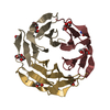
| Title | Cocrystal of an RSL-N23H and sulfonato-thiacalix[4]arene - zinc complex | ||||||||||||
 Components Components | Fucose-binding lectin protein | ||||||||||||
 Keywords Keywords | SUGAR BINDING PROTEIN /  calixarene / molecular glue / metal binding / synthetic receptor / b-propeller / thiacalixarene calixarene / molecular glue / metal binding / synthetic receptor / b-propeller / thiacalixarene | ||||||||||||
| Function / homology | Fucose-specific lectin / Fungal fucose-specific lectin / carbohydrate binding / sulfonato-thiacalix[4]arene / beta-D-fructopyranose / Fucose-binding lectin protein Function and homology information Function and homology information | ||||||||||||
| Biological species |  Ralstonia solanacearum (bacteria) Ralstonia solanacearum (bacteria) | ||||||||||||
| Method | X-RAY DIFFRACTION / SYNCHROTRON / MOLECULAR REPLACEMENT / molecular replacement / Resolution: 1.94 Å | ||||||||||||
 Authors Authors | Flood, R.J. / Ramberg, K. / Guagnini, F. / Crowley, P.B. | ||||||||||||
| Funding support |  Ireland, 3items Ireland, 3items
| ||||||||||||
 Citation Citation | Journal: Cryst.Growth Des. / Year: 2022 Title: Protein Frameworks with Thiacalixarene and Zinc. Authors: Flood, R.J. / Ramberg, K.O. / Mengel, D.B. / Guagnini, F. / Crowley, P.B. | ||||||||||||
| History |
|
- Structure visualization
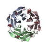
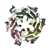
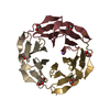
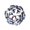
| Structure viewer | Molecule: MolmilJmol/JSmol |
|---|
- Downloads & links
-Download
| PDBx/mmCIF format | 7pr5.cif.gz | 241.9 KB | Display | PDBx/mmCIF format |
|---|---|---|---|---|
| PDB format | pdb7pr5.ent.gz | 194.3 KB | Display | PDB format |
| PDBx/mmJSON format | 7pr5.json.gz | Tree view | PDBx/mmJSON format | |
| Others |  Other downloads Other downloads |
-Validation report
| Arichive directory | https://data.pdbj.org/pub/pdb/validation_reports/pr/7pr5ftp://data.pdbj.org/pub/pdb/validation_reports/pr/7pr5 | HTTPS FTP |
|---|
-Related structure data
| Related structure data | 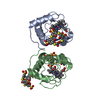 7pr2C 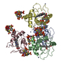 7pr3C  7pr4C 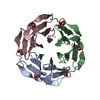 2bt9S C: citing same article ( S: Starting model for refinement |
|---|---|
| Similar structure data |


-Links
 PDBj
PDBj- Assembly
| Deposited unit | 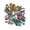
| ||||||||
|---|---|---|---|---|---|---|---|---|---|
| 1 | 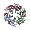
| ||||||||
| 2 | 
| ||||||||
| 3 | 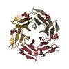
| ||||||||
| 4 | 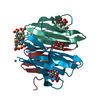
| ||||||||
| Unit cell |
|
-Components
-Protein / Sugars , 2 types, 35 molecules ABCDEFGHIJKL
| #1: Protein | Mass: 9757.606 Da / Num. of mol.: 12 Source method: isolated from a genetically manipulated source Source: (gene. exp.) Ralstonia solanacearum (bacteria)Gene: E7Z57_08365, HXP36_18875, RSP795_21825, RSP822_19650, RUN39_v1_50103 Production host: Escherichia coli (E. coli) / References: UniProt: A0A0S4TLR1#2: Sugar | ChemComp-BDF / Fructose Type: D-saccharide, beta linking / Mass: 180.156 Da / Num. of mol.: 23 / Source method: obtained synthetically / Formula: C6H12O6 Type: D-saccharide, beta linking / Mass: 180.156 Da / Num. of mol.: 23 / Source method: obtained synthetically / Formula: C6H12O6 |
|---|
-Non-polymers , 4 types, 867 molecules 


| #3: Chemical | ChemComp-ZN /  Mass: 65.409 Da / Num. of mol.: 7 / Source method: obtained synthetically / Formula: Zn Mass: 65.409 Da / Num. of mol.: 7 / Source method: obtained synthetically / Formula: Zn#4: Chemical | ChemComp-GOL / | Glycerol Mass: 92.094 Da / Num. of mol.: 1 / Source method: obtained synthetically / Formula: C3H8O3 Mass: 92.094 Da / Num. of mol.: 1 / Source method: obtained synthetically / Formula: C3H8O3#5: Chemical | ChemComp-80M / |  Mass: 816.894 Da / Num. of mol.: 1 / Source method: obtained synthetically / Formula: C24H16O16S8 / Feature type: SUBJECT OF INVESTIGATION Mass: 816.894 Da / Num. of mol.: 1 / Source method: obtained synthetically / Formula: C24H16O16S8 / Feature type: SUBJECT OF INVESTIGATION#6: Water | ChemComp-HOH / | WaterMass: 18.015 Da / Num. of mol.: 858 / Source method: isolated from a natural source / Formula: H2O |
|---|
-Details
| Has ligand of interest | Y |
|---|
-Experimental details
-Experiment
| Experiment | Method: X-RAY DIFFRACTION / Number of used crystals: 1 |
|---|
- Sample preparation
| Crystal | Density Matthews: 2.45 Å3/Da / Density % sol: 49.8 % |
|---|---|
| Crystal grow | Temperature: 293 K / Method: vapor diffusion, sitting drop / pH: 8.3 Details: 16% PEG 3350, 200 mM tri-Sodium citrate pH 8.3, 60 mM zinc acetate |
-Data collection
| Diffraction | Mean temperature: 100 K / Serial crystal experiment: N | ||||||||||||||||||||||||||||||
|---|---|---|---|---|---|---|---|---|---|---|---|---|---|---|---|---|---|---|---|---|---|---|---|---|---|---|---|---|---|---|---|
| Diffraction source | Source: SYNCHROTRON / Site: SOLEIL  / Beamline: PROXIMA 2 / Wavelength: 0.98 Å / Beamline: PROXIMA 2 / Wavelength: 0.98 Å | ||||||||||||||||||||||||||||||
| Detector | Type: DECTRIS EIGER X 9M / Detector: PIXEL / Date: Jul 25, 2020 | ||||||||||||||||||||||||||||||
| Radiation | Protocol: SINGLE WAVELENGTH / Monochromatic (M) / Laue (L): M / Scattering type: x-ray | ||||||||||||||||||||||||||||||
| Radiation wavelength | Wavelength: 0.98 Å / Relative weight: 1 | ||||||||||||||||||||||||||||||
| Reflection | Resolution: 1.94→46.84 Å / Num. obs: 85149 / % possible obs: 99.4 % / Redundancy: 13.6 % / Biso Wilson estimate: 32.44 Å2 / CC1/2: 0.999 / Rmerge(I) obs: 0.127 / Rpim(I) all: 0.036 / Rrim(I) all: 0.132 / Net I/σ(I): 14.8 / Num. measured all: 1155816 | ||||||||||||||||||||||||||||||
| Reflection shell | Diffraction-ID: 1
|
-Phasing
| Phasing | Method: molecular replacement | |||||||||
|---|---|---|---|---|---|---|---|---|---|---|
| Phasing MR | Model details: Phaser MODE: MR_AUTO
|
- Processing
| Software |
| |||||||||||||||||||||||||||||||||||||||||||||||||||||||||||||||||||||||||||||||||||||||||||||||||||||||||||||||||||||||||||||||||||||||||||||||||||||||||||||||||||||||||||||||||||||||||||||||||||||||||||||||||||||||||
|---|---|---|---|---|---|---|---|---|---|---|---|---|---|---|---|---|---|---|---|---|---|---|---|---|---|---|---|---|---|---|---|---|---|---|---|---|---|---|---|---|---|---|---|---|---|---|---|---|---|---|---|---|---|---|---|---|---|---|---|---|---|---|---|---|---|---|---|---|---|---|---|---|---|---|---|---|---|---|---|---|---|---|---|---|---|---|---|---|---|---|---|---|---|---|---|---|---|---|---|---|---|---|---|---|---|---|---|---|---|---|---|---|---|---|---|---|---|---|---|---|---|---|---|---|---|---|---|---|---|---|---|---|---|---|---|---|---|---|---|---|---|---|---|---|---|---|---|---|---|---|---|---|---|---|---|---|---|---|---|---|---|---|---|---|---|---|---|---|---|---|---|---|---|---|---|---|---|---|---|---|---|---|---|---|---|---|---|---|---|---|---|---|---|---|---|---|---|---|---|---|---|---|---|---|---|---|---|---|---|---|---|---|---|---|---|---|---|---|
| Refinement | Method to determine structure: MOLECULAR REPLACEMENT Starting model: 2BT9 Resolution: 1.94→46.84 Å / SU ML: 0.27 / Cross valid method: THROUGHOUT / σ(F): 1.34 / Phase error: 22.99 / Stereochemistry target values: ML
| |||||||||||||||||||||||||||||||||||||||||||||||||||||||||||||||||||||||||||||||||||||||||||||||||||||||||||||||||||||||||||||||||||||||||||||||||||||||||||||||||||||||||||||||||||||||||||||||||||||||||||||||||||||||||
| Solvent computation | Shrinkage radii: 0.9 Å / VDW probe radii: 1.11 Å / Solvent model: FLAT BULK SOLVENT MODEL | |||||||||||||||||||||||||||||||||||||||||||||||||||||||||||||||||||||||||||||||||||||||||||||||||||||||||||||||||||||||||||||||||||||||||||||||||||||||||||||||||||||||||||||||||||||||||||||||||||||||||||||||||||||||||
| Displacement parameters | Biso max: 76.6 Å2 / Biso mean: 33.5422 Å2 / Biso min: 17.46 Å2 | |||||||||||||||||||||||||||||||||||||||||||||||||||||||||||||||||||||||||||||||||||||||||||||||||||||||||||||||||||||||||||||||||||||||||||||||||||||||||||||||||||||||||||||||||||||||||||||||||||||||||||||||||||||||||
| Refinement step | Cycle: final / Resolution: 1.94→46.84 Å
| |||||||||||||||||||||||||||||||||||||||||||||||||||||||||||||||||||||||||||||||||||||||||||||||||||||||||||||||||||||||||||||||||||||||||||||||||||||||||||||||||||||||||||||||||||||||||||||||||||||||||||||||||||||||||
| LS refinement shell | Refine-ID: X-RAY DIFFRACTION / Rfactor Rfree error: 0 / Total num. of bins used: 30
|
