 Movie
Movie Controller

[English] 日本語
 Yorodumi
- PDB-7pq2: Crystal Structure of the Ring Nuclease 0811 from Sulfolobus islan... -
+ Open data

- Basic information
| Entry | Database: PDB / ID: 7pq2 | ||||||
|---|---|---|---|---|---|---|---|
| Title | Crystal Structure of the Ring Nuclease 0811 from Sulfolobus islandicus (Sis0811) in its apo form | ||||||
 Components Components | CRISPR-associated protein, APE2256 family, CRISPR Ring Nuclease | ||||||
 Keywords Keywords |  ANTIVIRAL PROTEIN / RING NUCLEASE / CRISPR-ASSOCIATED PROTEIN / HELIX-TURN-HELIX / VIRAL RESISTANCE / CARF NUCLEOTIDE-BINDING DOMAIN / CRISPR Ring Nuclease ANTIVIRAL PROTEIN / RING NUCLEASE / CRISPR-ASSOCIATED PROTEIN / HELIX-TURN-HELIX / VIRAL RESISTANCE / CARF NUCLEOTIDE-BINDING DOMAIN / CRISPR Ring Nuclease | ||||||
| Function / homology | CRISPR system ring nuclease SSO1393 / CRISPR system ring nuclease SSO1393-like / CRISPR-associated protein (Cas_APE2256) / CRISPR-associated protein, APE2256 family Function and homology information Function and homology information | ||||||
| Biological species |   Sulfolobus islandicus REY15A (acidophilic) Sulfolobus islandicus REY15A (acidophilic) | ||||||
| Method | X-RAY DIFFRACTION / SYNCHROTRON / MOLECULAR REPLACEMENT / Resolution: 2.38 Å | ||||||
 Authors Authors | Molina, R. / Jensen, A.L.G. / Marchena-Hurtado, J. / Lopez-Mendez, B. / Stella, S. / Montoya, G. | ||||||
| Funding support |  Denmark, 1items Denmark, 1items
| ||||||
 Citation Citation | Journal: Nucleic Acids Res. / Year: 2021 Title: Structural basis of cyclic oligoadenylate degradation by ancillary Type III CRISPR-Cas ring nucleases. Authors: Molina, R. / Jensen, A.L.G. / Marchena-Hurtado, J. / Lopez-Mendez, B. / Stella, S. / Montoya, G. | ||||||
| History |
|
- Structure visualization
| Structure viewer | Molecule: MolmilJmol/JSmol |
|---|
- Downloads & links
-Download
| PDBx/mmCIF format | 7pq2.cif.gz | 215.9 KB | Display | PDBx/mmCIF format |
|---|---|---|---|---|
| PDB format | pdb7pq2.ent.gz | Display | PDB format | |
| PDBx/mmJSON format | 7pq2.json.gz | Tree view | PDBx/mmJSON format | |
| Others |  Other downloads Other downloads |
-Validation report
| Arichive directory | https://data.pdbj.org/pub/pdb/validation_reports/pq/7pq2ftp://data.pdbj.org/pub/pdb/validation_reports/pq/7pq2 | HTTPS FTP |
|---|
-Related structure data
| Related structure data | 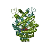 7pq3C  7pq6C  7pqaC  3qyfS S: Starting model for refinement C: citing same article ( |
|---|---|
| Similar structure data |


-Links
 PDBj
PDBj- Assembly
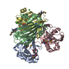
| Deposited unit | 
| ||||||||
|---|---|---|---|---|---|---|---|---|---|
| 1 |
| ||||||||
| Unit cell |
| ||||||||
| Noncrystallographic symmetry (NCS) | NCS domain: (Details: Chains AAA BBB) |
-Components
| #1: Protein | Mass: 31654.133 Da / Num. of mol.: 2 Source method: isolated from a genetically manipulated source Source: (gene. exp.) Sulfolobus islandicus REY15A (acidophilic)Gene: SiRe_0811 / Production host:  Escherichia coli BL21 (bacteria) / References: UniProt: F0NH89 Escherichia coli BL21 (bacteria) / References: UniProt: F0NH89#2: Water | ChemComp-HOH / | Water Mass: 18.015 Da / Num. of mol.: 254 / Source method: isolated from a natural source / Formula: H2O Mass: 18.015 Da / Num. of mol.: 254 / Source method: isolated from a natural source / Formula: H2O |
|---|
-Experimental details
-Experiment
| Experiment | Method: X-RAY DIFFRACTION / Number of used crystals: 1 |
|---|
- Sample preparation
| Crystal | Density Matthews: 2.49 Å3/Da / Density % sol: 50.53 % |
|---|---|
| Crystal grow | Temperature: 293 K / Method: vapor diffusion, hanging drop / Details: 30% PEG3000, 0.1M CHES pH 9.5 |
-Data collection
| Diffraction | Mean temperature: 100 K / Serial crystal experiment: N |
|---|---|
| Diffraction source | Source: SYNCHROTRON / Site: MAX IV  / Beamline: BioMAX / Wavelength: 0.98 Å / Beamline: BioMAX / Wavelength: 0.98 Å |
| Detector | Type: DECTRIS EIGER X 16M / Detector: PIXEL / Date: Nov 28, 2019 |
| Radiation | Protocol: SINGLE WAVELENGTH / Monochromatic (M) / Laue (L): M / Scattering type: x-ray |
| Radiation wavelength | Wavelength: 0.98 Å / Relative weight: 1 |
| Reflection | Resolution: 2.38→61.73 Å / Num. obs: 24724 / % possible obs: 99.9 % / Redundancy: 7 % / CC1/2: 0.99 / Net I/σ(I): 14.1 |
| Reflection shell | Resolution: 2.38→2.43 Å / Num. unique obs: 1241 / CC1/2: 0.756 |
- Processing
| Software |
| |||||||||||||||||||||||||||||||||||||||||||||||||||||||||||||||||||||||||||||||||||||||||||||||||||||||||||||||||||||||||||||||||||||||||||||||||||
|---|---|---|---|---|---|---|---|---|---|---|---|---|---|---|---|---|---|---|---|---|---|---|---|---|---|---|---|---|---|---|---|---|---|---|---|---|---|---|---|---|---|---|---|---|---|---|---|---|---|---|---|---|---|---|---|---|---|---|---|---|---|---|---|---|---|---|---|---|---|---|---|---|---|---|---|---|---|---|---|---|---|---|---|---|---|---|---|---|---|---|---|---|---|---|---|---|---|---|---|---|---|---|---|---|---|---|---|---|---|---|---|---|---|---|---|---|---|---|---|---|---|---|---|---|---|---|---|---|---|---|---|---|---|---|---|---|---|---|---|---|---|---|---|---|---|---|---|---|
| Refinement | Method to determine structure: MOLECULAR REPLACEMENT Starting model: 3QYF Resolution: 2.38→61.61 Å / Cor.coef. Fo:Fc: 0.963 / Cor.coef. Fo:Fc free: 0.933 / SU B: 15.932 / SU ML: 0.182 / Cross valid method: FREE R-VALUE / ESU R: 0.454 / ESU R Free: 0.25 Details: Hydrogens have been added in their riding positions
| |||||||||||||||||||||||||||||||||||||||||||||||||||||||||||||||||||||||||||||||||||||||||||||||||||||||||||||||||||||||||||||||||||||||||||||||||||
| Solvent computation | Ion probe radii: 0.8 Å / Shrinkage radii: 0.8 Å / VDW probe radii: 1.2 Å / Solvent model: MASK BULK SOLVENT | |||||||||||||||||||||||||||||||||||||||||||||||||||||||||||||||||||||||||||||||||||||||||||||||||||||||||||||||||||||||||||||||||||||||||||||||||||
| Displacement parameters | Biso mean: 42.532 Å2
| |||||||||||||||||||||||||||||||||||||||||||||||||||||||||||||||||||||||||||||||||||||||||||||||||||||||||||||||||||||||||||||||||||||||||||||||||||
| Refinement step | Cycle: LAST / Resolution: 2.38→61.61 Å
| |||||||||||||||||||||||||||||||||||||||||||||||||||||||||||||||||||||||||||||||||||||||||||||||||||||||||||||||||||||||||||||||||||||||||||||||||||
| Refine LS restraints |
| |||||||||||||||||||||||||||||||||||||||||||||||||||||||||||||||||||||||||||||||||||||||||||||||||||||||||||||||||||||||||||||||||||||||||||||||||||
| LS refinement shell |
| |||||||||||||||||||||||||||||||||||||||||||||||||||||||||||||||||||||||||||||||||||||||||||||||||||||||||||||||||||||||||||||||||||||||||||||||||||
| Refinement TLS params. | Method: refined / Refine-ID: X-RAY DIFFRACTION
| |||||||||||||||||||||||||||||||||||||||||||||||||||||||||||||||||||||||||||||||||||||||||||||||||||||||||||||||||||||||||||||||||||||||||||||||||||
| Refinement TLS group |
|
