 Movie
Movie Controller


+ Open data

- Basic information
| Entry | Database: PDB / ID: 7p57 | ||||||
|---|---|---|---|---|---|---|---|
| Title | VSG2 mutant structure lacking the calcium binding pocket | ||||||
 Components Components | Variant surface glycoprotein MITAT 1.2 | ||||||
 Keywords Keywords |  MEMBRANE PROTEIN / Variant Surface Glycoprotein / Coat / Trypanosoma brucei / African trypanosome / Immune Evasion / antigenic variation / calcium binding / calcium MEMBRANE PROTEIN / Variant Surface Glycoprotein / Coat / Trypanosoma brucei / African trypanosome / Immune Evasion / antigenic variation / calcium binding / calcium | ||||||
| Function / homology | Variant surface glycoprotein C-terminal domain superfamily / Trypanosome variant surface glycoprotein, A-type, N-terminal domain / Trypanosome variant surface glycoprotein (A-type) / evasion of host immune response / side of membrane / plasma membrane / Variant surface glycoprotein MITAT 1.2 Function and homology information Function and homology information | ||||||
| Biological species |  Trypanosoma brucei brucei (eukaryote) Trypanosoma brucei brucei (eukaryote) | ||||||
| Method | X-RAY DIFFRACTION / SYNCHROTRON / MOLECULAR REPLACEMENT / Resolution: 1.961 Å | ||||||
 Authors Authors | Gkeka, A. / Aresta-Branco, F. / Stebbins, C.E. / Papavasiliou, F.N. | ||||||
 Citation Citation | Journal: Cell Rep / Year: 2023 Title: Immunodominant surface epitopes power immune evasion in the African trypanosome. Authors: Gkeka, A. / Aresta-Branco, F. / Triller, G. / Vlachou, E.P. / van Straaten, M. / Lilic, M. / Olinares, P.D.B. / Perez, K. / Chait, B.T. / Blatnik, R. / Ruppert, T. / Verdi, J.P. / Stebbins, ...Authors: Gkeka, A. / Aresta-Branco, F. / Triller, G. / Vlachou, E.P. / van Straaten, M. / Lilic, M. / Olinares, P.D.B. / Perez, K. / Chait, B.T. / Blatnik, R. / Ruppert, T. / Verdi, J.P. / Stebbins, C.E. / Papavasiliou, F.N. #1: Journal: Biorxiv / Year: 2022Title: Immunodominant surface epitopes power immune evasion in the African trypanosome Authors: Gkeka, A. / Aresta-Branco, F. / Triller, G. / Vlachou, E.P. / Lilic, M. / Olinares, P.D.B. / Perez, K. / Chait, B.T. / Blatnik, R. / Ruppert, T. / Stebbins, C.E. / Papavasiliou, F.N. | ||||||
| History |
|
- Structure visualization
| Structure viewer | Molecule: MolmilJmol/JSmol |
|---|
- Downloads & links
-Download
| PDBx/mmCIF format | 7p57.cif.gz | 160 KB | Display | PDBx/mmCIF format |
|---|---|---|---|---|
| PDB format | pdb7p57.ent.gz | 123.5 KB | Display | PDB format |
| PDBx/mmJSON format | 7p57.json.gz | Tree view | PDBx/mmJSON format | |
| Others |  Other downloads Other downloads |
-Validation report
| Arichive directory | https://data.pdbj.org/pub/pdb/validation_reports/p5/7p57ftp://data.pdbj.org/pub/pdb/validation_reports/p5/7p57 | HTTPS FTP |
|---|
-Related structure data
| Related structure data |  7p56C 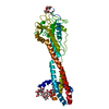 7p59C  7p5aC  7p5bC 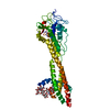 7p5dC  1vsgS S: Starting model for refinement C: citing same article ( |
|---|---|
| Similar structure data |
-Links
 PDBj
PDBj- Assembly
| Deposited unit | 
| ||||||||
|---|---|---|---|---|---|---|---|---|---|
| 1 |
| ||||||||
| 2 | 
| ||||||||
| Unit cell |
| ||||||||
| Components on special symmetry positions |
|
-Components
| #1: Protein | Mass: 50971.387 Da / Num. of mol.: 1 Source method: isolated from a genetically manipulated source Details: VSG2 "AAA mutant" (D208, N209, and D210 each mutated to alanine). Source: (gene. exp.) Trypanosoma brucei brucei (eukaryote) / Cell line (production host): FAB 3.6.4 (VSG2-AAA) / Production host: Trypanosoma brucei brucei (eukaryote) / References: UniProt: P26332 |
|---|---|
| #2: Polysaccharide | alpha-D-mannopyranose-(1-3)-beta-D-mannopyranose-(1-4)-2-acetamido-2-deoxy-beta-D-glucopyranose-(1- ...alpha-D-mannopyranose-(1-3)-beta-D-mannopyranose-(1-4)-2-acetamido-2-deoxy-beta-D-glucopyranose-(1-4)-2-acetamido-2-deoxy-beta-D-glucopyranose / Mass: 748.682 Da / Num. of mol.: 1 Source method: isolated from a genetically manipulated source |
| #3: Water | ChemComp-HOH / Water Mass: 18.015 Da / Num. of mol.: 239 / Source method: isolated from a natural source / Formula: H2O Mass: 18.015 Da / Num. of mol.: 239 / Source method: isolated from a natural source / Formula: H2O |
| Has ligand of interest | N |
-Experimental details
-Experiment
| Experiment | Method: X-RAY DIFFRACTION / Number of used crystals: 1 |
|---|
- Sample preparation
| Crystal | Density Matthews: 2.45 Å3/Da / Density % sol: 49.82 % |
|---|---|
| Crystal grow | Temperature: 295 K / Method: vapor diffusion, hanging drop / Details: 0.1M Tris pH 8.0, 39% PEG400 |
-Data collection
| Diffraction | Mean temperature: 100 K / Serial crystal experiment: N |
|---|---|
| Diffraction source | Source: SYNCHROTRON / Site: SLS  / Beamline: X06DA / Wavelength: 1 Å / Beamline: X06DA / Wavelength: 1 Å |
| Detector | Type: DECTRIS PILATUS 2M-F / Detector: PIXEL / Date: Oct 27, 2019 |
| Radiation | Protocol: SINGLE WAVELENGTH / Monochromatic (M) / Laue (L): M / Scattering type: x-ray |
| Radiation wavelength | Wavelength: 1 Å / Relative weight: 1 |
| Reflection | Resolution: 1.961→48.01 Å / Num. obs: 36675 / % possible obs: 99.24 % / Redundancy: 19.7 % / Biso Wilson estimate: 34.2 Å2 / CC1/2: 0.999 / CC star: 1 / Rmerge(I) obs: 0.09816 / Rpim(I) all: 0.02123 / Rrim(I) all: 0.1006 / Net I/σ(I): 20.27 |
| Reflection shell | Resolution: 1.961→2.031 Å / Redundancy: 3.6 % / Rmerge(I) obs: 0.8934 / Mean I/σ(I) obs: 0.84 / Num. unique obs: 3341 / CC1/2: 0.557 / CC star: 0.846 / Rpim(I) all: 0.4747 / Rrim(I) all: 1.023 / % possible all: 92.39 |
- Processing
| Software |
| |||||||||||||||||||||||||||||||||||||||||||||||||||||||||||||||||||||||||||||||||||||||||||||||||||||||||||||||||||||||||||||||||||||||||||||||||||||||||||||||||||||||||||||||||||||||||||||||||||||||||||||||||||||||||||||||||||||||||||||||||||||||||||||||||||||||||||||||||||
|---|---|---|---|---|---|---|---|---|---|---|---|---|---|---|---|---|---|---|---|---|---|---|---|---|---|---|---|---|---|---|---|---|---|---|---|---|---|---|---|---|---|---|---|---|---|---|---|---|---|---|---|---|---|---|---|---|---|---|---|---|---|---|---|---|---|---|---|---|---|---|---|---|---|---|---|---|---|---|---|---|---|---|---|---|---|---|---|---|---|---|---|---|---|---|---|---|---|---|---|---|---|---|---|---|---|---|---|---|---|---|---|---|---|---|---|---|---|---|---|---|---|---|---|---|---|---|---|---|---|---|---|---|---|---|---|---|---|---|---|---|---|---|---|---|---|---|---|---|---|---|---|---|---|---|---|---|---|---|---|---|---|---|---|---|---|---|---|---|---|---|---|---|---|---|---|---|---|---|---|---|---|---|---|---|---|---|---|---|---|---|---|---|---|---|---|---|---|---|---|---|---|---|---|---|---|---|---|---|---|---|---|---|---|---|---|---|---|---|---|---|---|---|---|---|---|---|---|---|---|---|---|---|---|---|---|---|---|---|---|---|---|---|---|---|---|---|---|---|---|---|---|---|---|---|---|---|---|---|---|---|---|---|---|---|---|---|---|---|---|---|---|---|---|---|---|---|
| Refinement | Method to determine structure: MOLECULAR REPLACEMENT Starting model: 1VSG Resolution: 1.961→48.01 Å / SU ML: 0.2 / Cross valid method: FREE R-VALUE / σ(F): 1.37 / Phase error: 19.32 / Stereochemistry target values: ML
| |||||||||||||||||||||||||||||||||||||||||||||||||||||||||||||||||||||||||||||||||||||||||||||||||||||||||||||||||||||||||||||||||||||||||||||||||||||||||||||||||||||||||||||||||||||||||||||||||||||||||||||||||||||||||||||||||||||||||||||||||||||||||||||||||||||||||||||||||||
| Solvent computation | Shrinkage radii: 0.9 Å / VDW probe radii: 1.11 Å / Solvent model: FLAT BULK SOLVENT MODEL | |||||||||||||||||||||||||||||||||||||||||||||||||||||||||||||||||||||||||||||||||||||||||||||||||||||||||||||||||||||||||||||||||||||||||||||||||||||||||||||||||||||||||||||||||||||||||||||||||||||||||||||||||||||||||||||||||||||||||||||||||||||||||||||||||||||||||||||||||||
| Displacement parameters | Biso max: 105.86 Å2 / Biso mean: 43.8988 Å2 / Biso min: 21.36 Å2 | |||||||||||||||||||||||||||||||||||||||||||||||||||||||||||||||||||||||||||||||||||||||||||||||||||||||||||||||||||||||||||||||||||||||||||||||||||||||||||||||||||||||||||||||||||||||||||||||||||||||||||||||||||||||||||||||||||||||||||||||||||||||||||||||||||||||||||||||||||
| Refinement step | Cycle: final / Resolution: 1.961→48.01 Å
| |||||||||||||||||||||||||||||||||||||||||||||||||||||||||||||||||||||||||||||||||||||||||||||||||||||||||||||||||||||||||||||||||||||||||||||||||||||||||||||||||||||||||||||||||||||||||||||||||||||||||||||||||||||||||||||||||||||||||||||||||||||||||||||||||||||||||||||||||||
| LS refinement shell | Refine-ID: X-RAY DIFFRACTION / Rfactor Rfree error: 0
| |||||||||||||||||||||||||||||||||||||||||||||||||||||||||||||||||||||||||||||||||||||||||||||||||||||||||||||||||||||||||||||||||||||||||||||||||||||||||||||||||||||||||||||||||||||||||||||||||||||||||||||||||||||||||||||||||||||||||||||||||||||||||||||||||||||||||||||||||||
| Refinement TLS params. | Method: refined / Refine-ID: X-RAY DIFFRACTION
| |||||||||||||||||||||||||||||||||||||||||||||||||||||||||||||||||||||||||||||||||||||||||||||||||||||||||||||||||||||||||||||||||||||||||||||||||||||||||||||||||||||||||||||||||||||||||||||||||||||||||||||||||||||||||||||||||||||||||||||||||||||||||||||||||||||||||||||||||||
| Refinement TLS group |
|
