 Movie
Movie Controller

[English] 日本語
 Yorodumi
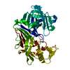
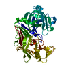
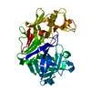
- PDB-7oyu: E.coli's putrescine receptor variant PotF/D (4JDF) with mutations... -
+ Open data

- Basic information
| Entry | Database: PDB / ID: 7oyu | ||||||
|---|---|---|---|---|---|---|---|
| Title | E.coli's putrescine receptor variant PotF/D (4JDF) with mutations E39D Y87S F88Y in complex with spermidine | ||||||
 Components Components | Putrescine-binding periplasmic protein PotF | ||||||
 Keywords Keywords |  TRANSPORT PROTEIN / E.coli / Periplasmic binding protein / PotF / Spermidine TRANSPORT PROTEIN / E.coli / Periplasmic binding protein / PotF / Spermidine | ||||||
| Function / homology |  Function and homology information Function and homology informationputrescine transport / putrescine binding / ATP-binding cassette (ABC) transporter complex, substrate-binding subunit-containing / outer membrane-bounded periplasmic space / membraneSimilarity search - Function | ||||||
| Biological species |  Escherichia coli (E. coli) Escherichia coli (E. coli) | ||||||
| Method | X-RAY DIFFRACTION / SYNCHROTRON / MOLECULAR REPLACEMENT / Resolution: 1.95 Å | ||||||
 Authors Authors | Shanmugaratnam, S. / Kroeger, P. / Hocker, B. | ||||||
| Funding support |  Germany, 1items Germany, 1items
| ||||||
 Citation Citation | Journal: J.Biol.Chem. / Year: 2021 Title: Fine-tuning spermidine binding modes in the putrescine binding protein PotF. Authors: Kroger, P. / Shanmugaratnam, S. / Scheib, U. / Hocker, B. | ||||||
| History |
|
- Structure visualization
| Structure viewer | Molecule: MolmilJmol/JSmol |
|---|
- Downloads & links
-Download
| PDBx/mmCIF format | 7oyu.cif.gz | 184.8 KB | Display | PDBx/mmCIF format |
|---|---|---|---|---|
| PDB format | pdb7oyu.ent.gz | 120.8 KB | Display | PDB format |
| PDBx/mmJSON format | 7oyu.json.gz | Tree view | PDBx/mmJSON format | |
| Others |  Other downloads Other downloads |
-Validation report
| Arichive directory | https://data.pdbj.org/pub/pdb/validation_reports/oy/7oyuftp://data.pdbj.org/pub/pdb/validation_reports/oy/7oyu | HTTPS FTP |
|---|
-Related structure data
| Related structure data |  7oysC  7oytC  7oyvC  7oywC  7oyxC  7oyyC  7oyzC  1a99S S: Starting model for refinement C: citing same article ( |
|---|---|
| Similar structure data |


-Links
 PDBj
PDBj


- Assembly
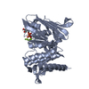
| Deposited unit | 
| ||||||||||||
|---|---|---|---|---|---|---|---|---|---|---|---|---|---|
| 1 |
| ||||||||||||
| Unit cell |
|
-Components
-Protein , 1 types, 1 molecules A
| #1: Protein | Mass: 39470.566 Da / Num. of mol.: 1 / Mutation: S38T, F88Y, A182D, D247S, F276W, L348Q Source method: isolated from a genetically manipulated source Source: (gene. exp.) Escherichia coli (strain K12) (bacteria)Strain: K12 / Gene: potF, b0854, JW0838 / Plasmid: pET21b(+) / Production host: Escherichia coli BL21(DE3) (bacteria) / References: UniProt: P31133 |
|---|
-Non-polymers , 5 types, 190 molecules 


| #2: Chemical | ChemComp-PGE / Polyethylene glycol Mass: 150.173 Da / Num. of mol.: 1 / Source method: obtained synthetically / Formula: C6H14O4 Mass: 150.173 Da / Num. of mol.: 1 / Source method: obtained synthetically / Formula: C6H14O4 | ||||||
|---|---|---|---|---|---|---|---|
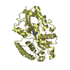
| #3: Chemical | ChemComp-EDO / Ethylene glycol Mass: 62.068 Da / Num. of mol.: 7 / Source method: obtained synthetically / Formula: C2H6O2 Mass: 62.068 Da / Num. of mol.: 7 / Source method: obtained synthetically / Formula: C2H6O2#4: Chemical | ChemComp-PEG / | Diethylene glycol Mass: 106.120 Da / Num. of mol.: 1 / Source method: obtained synthetically / Formula: C4H10O3 Mass: 106.120 Da / Num. of mol.: 1 / Source method: obtained synthetically / Formula: C4H10O3#5: Chemical | ChemComp-SPD / | Spermidine Mass: 145.246 Da / Num. of mol.: 1 / Source method: obtained synthetically / Formula: C7H19N3 / Feature type: SUBJECT OF INVESTIGATION Mass: 145.246 Da / Num. of mol.: 1 / Source method: obtained synthetically / Formula: C7H19N3 / Feature type: SUBJECT OF INVESTIGATION#6: Water | ChemComp-HOH / | WaterMass: 18.015 Da / Num. of mol.: 180 / Source method: isolated from a natural source / Formula: H2O |
-Details
| Has ligand of interest | Y |
|---|
-Experimental details
-Experiment
| Experiment | Method: X-RAY DIFFRACTION / Number of used crystals: 1 |
|---|
- Sample preparation
| Crystal | Density Matthews: 2.14 Å3/Da / Density % sol: 42.07 % |
|---|---|
| Crystal grow | Temperature: 293 K / Method: vapor diffusion, sitting drop / pH: 4.6 Details: 0.1 M Sodium acetate pH 4.6, 0.2 M Ammonium acetate, 30% PEG 4000 |
-Data collection
| Diffraction | Mean temperature: 100 K / Serial crystal experiment: N |
|---|---|
| Diffraction source | Source: SYNCHROTRON / Site: BESSY / Beamline: 14.2 / Wavelength: 0.9184 Å |
| Detector | Type: DECTRIS PILATUS3 2M / Detector: PIXEL / Date: Jul 11, 2019 |
| Radiation | Monochromator: DCM Si(111) / Protocol: SINGLE WAVELENGTH / Monochromatic (M) / Laue (L): M / Scattering type: x-ray |
| Radiation wavelength | Wavelength: 0.9184 Å / Relative weight: 1 |
| Reflection | Resolution: 1.95→46.36 Å / Num. obs: 25021 / % possible obs: 98.9 % / Redundancy: 5.5 % / Biso Wilson estimate: 30.12 Å2 / CC1/2: 0.994 / CC star: 0.998 / Rmerge(I) obs: 0.206 / Rpim(I) all: 0.093 / Rrim(I) all: 0.227 / Net I/σ(I): 6.44 |
| Reflection shell | Resolution: 1.95→2.02 Å / Redundancy: 5.7 % / Rmerge(I) obs: 2.231 / Mean I/σ(I) obs: 0.79 / Num. unique obs: 14023 / CC1/2: 0.262 / CC star: 0.644 / Rpim(I) all: 0.996 / Rrim(I) all: 2.451 / % possible all: 99.2 |
- Processing
| Software |
| ||||||||||||||||||||||||||||||||||||||||||||||||||||||||||||||||||||||
|---|---|---|---|---|---|---|---|---|---|---|---|---|---|---|---|---|---|---|---|---|---|---|---|---|---|---|---|---|---|---|---|---|---|---|---|---|---|---|---|---|---|---|---|---|---|---|---|---|---|---|---|---|---|---|---|---|---|---|---|---|---|---|---|---|---|---|---|---|---|---|---|
| Refinement | Method to determine structure: MOLECULAR REPLACEMENT Starting model: 1A99 Resolution: 1.95→46.36 Å / SU ML: 0.2521 / Cross valid method: FREE R-VALUE / Phase error: 25.0649 Stereochemistry target values: GeoStd + Monomer Library + CDL v1.2
| ||||||||||||||||||||||||||||||||||||||||||||||||||||||||||||||||||||||
| Solvent computation | Shrinkage radii: 0.9 Å / VDW probe radii: 1.11 Å / Solvent model: FLAT BULK SOLVENT MODEL | ||||||||||||||||||||||||||||||||||||||||||||||||||||||||||||||||||||||
| Displacement parameters | Biso mean: 35.92 Å2 | ||||||||||||||||||||||||||||||||||||||||||||||||||||||||||||||||||||||
| Refinement step | Cycle: LAST / Resolution: 1.95→46.36 Å
| ||||||||||||||||||||||||||||||||||||||||||||||||||||||||||||||||||||||
| Refine LS restraints |
| ||||||||||||||||||||||||||||||||||||||||||||||||||||||||||||||||||||||
| LS refinement shell |
| ||||||||||||||||||||||||||||||||||||||||||||||||||||||||||||||||||||||
| Refinement TLS params. | Method: refined / Origin x: -0.328129966486 Å / Origin y: -0.0287458903317 Å / Origin z: -11.9408554634 Å
| ||||||||||||||||||||||||||||||||||||||||||||||||||||||||||||||||||||||
| Refinement TLS group | Selection details: (chain 'A' and resid 29 through 369) |
