 Movie
Movie Controller

[English] 日本語
 Yorodumi
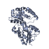
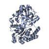
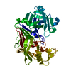
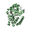
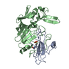
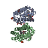
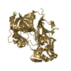
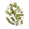
- PDB-3ttn: Crystal structures of polyamine receptors SpuD and SpuE from Pseu... -
+ Open data

- Basic information
| Entry | Database: PDB / ID: 3ttn | ||||||
|---|---|---|---|---|---|---|---|
| Title | Crystal structures of polyamine receptors SpuD and SpuE from Pseudomonas aeruginosa | ||||||
 Components Components | Polyamine transport protein | ||||||
 Keywords Keywords |  TRANSPORT PROTEIN / polyamine binding protein / spermidine TRANSPORT PROTEIN / polyamine binding protein / spermidine | ||||||
| Function / homology |  Function and homology information Function and homology informationpolyamine transport / spermidine binding / periplasmic space / extracellular spaceSimilarity search - Function | ||||||
| Biological species |   Pseudomonas aeruginosa (bacteria) Pseudomonas aeruginosa (bacteria) | ||||||
| Method | X-RAY DIFFRACTION / SYNCHROTRON / SAD / Resolution: 2 Å | ||||||
 Authors Authors | Lim, S.C. / Wu, D.H. / Song, H.W. | ||||||
 Citation Citation | Journal: J.Mol.Biol. / Year: 2012 Title: Structural Basis of Substrate Binding Specificity Revealed by the Crystal Structures of Polyamine Receptors SpuD and SpuE from Pseudomonas aeruginosa Authors: Wu, D.H. / Lim, S.C. / Dong, Y.H. / Wu, J.E. / Tao, F. / Zhou, L. / Zhang, L.H. / Song, H.W. | ||||||
| History |
|
- Structure visualization
| Structure viewer | Molecule: MolmilJmol/JSmol |
|---|
- Downloads & links
-Download
| PDBx/mmCIF format | 3ttn.cif.gz | 137.7 KB | Display | PDBx/mmCIF format |
|---|---|---|---|---|
| PDB format | pdb3ttn.ent.gz | 113.7 KB | Display | PDB format |
| PDBx/mmJSON format | 3ttn.json.gz | Tree view | PDBx/mmJSON format | |
| Others |  Other downloads Other downloads |
-Validation report
| Arichive directory | https://data.pdbj.org/pub/pdb/validation_reports/tt/3ttnftp://data.pdbj.org/pub/pdb/validation_reports/tt/3ttn | HTTPS FTP |
|---|
-Related structure data


-Links
 PDBj
PDBj


- Assembly
| Deposited unit | 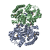
| |||||||||||||||||||||||||||
|---|---|---|---|---|---|---|---|---|---|---|---|---|---|---|---|---|---|---|---|---|---|---|---|---|---|---|---|---|
| 1 | 
| |||||||||||||||||||||||||||
| 2 | 
| |||||||||||||||||||||||||||
| Unit cell |
| |||||||||||||||||||||||||||
| Noncrystallographic symmetry (NCS) | NCS domain:
NCS domain segments:
|
-Components
| #1: Protein | Mass: 37737.875 Da / Num. of mol.: 2 / Fragment: UNP residues 28-362 Source method: isolated from a genetically manipulated source Source: (gene. exp.) Pseudomonas aeruginosa (bacteria) / Gene: spuE / Production host: Escherichia coli (E. coli) / References: UniProt: Q9I6J0#2: Chemical | Spermidine  Mass: 145.246 Da / Num. of mol.: 2 / Source method: obtained synthetically / Formula: C7H19N3 Mass: 145.246 Da / Num. of mol.: 2 / Source method: obtained synthetically / Formula: C7H19N3#3: Water | ChemComp-HOH / | Water Mass: 18.015 Da / Num. of mol.: 197 / Source method: isolated from a natural source / Formula: H2O Mass: 18.015 Da / Num. of mol.: 197 / Source method: isolated from a natural source / Formula: H2O |
|---|
-Experimental details
-Experiment
| Experiment | Method: X-RAY DIFFRACTION / Number of used crystals: 1 |
|---|
- Sample preparation
| Crystal | Density Matthews: 2.11 Å3/Da / Density % sol: 41.6 % |
|---|---|
| Crystal grow | Temperature: 288 K / Method: vapor diffusion, hanging drop / pH: 2.6 Details: 0.1M citric acid, 28% PEG 6000, 4% Jeffamine M-600, pH 2.6, VAPOR DIFFUSION, HANGING DROP, temperature 288K |
-Data collection
| Diffraction | Mean temperature: 100 K |
|---|---|
| Diffraction source | Source: SYNCHROTRON / Site: Australian Synchrotron  / Beamline: MX1 / Wavelength: 0.97988 Å / Beamline: MX1 / Wavelength: 0.97988 Å |
| Detector | Type: ADSC QUANTUM 210r / Detector: CCD / Date: Feb 25, 2009 |
| Radiation | Monochromator: Si 111 CHANNEL / Protocol: SINGLE WAVELENGTH / Monochromatic (M) / Laue (L): M / Scattering type: x-ray |
| Radiation wavelength | Wavelength: 0.97988 Å / Relative weight: 1 |
| Reflection | Resolution: 2→24.07 Å / Num. all: 40146 / Num. obs: 40146 / % possible obs: 96.5 % / Observed criterion σ(F): 2 / Observed criterion σ(I): 2 |
| Reflection shell | Resolution: 2→2.11 Å / % possible all: 87.3 |
- Processing
| Software |
| ||||||||||||||||||||||||||||||||||||||||||||||||||||||||||||||||||||||||||||||||||||||||||||||||||||||||||||||||||||||||||||||||||||||||||||||||||||||||||||||||||||||||||
|---|---|---|---|---|---|---|---|---|---|---|---|---|---|---|---|---|---|---|---|---|---|---|---|---|---|---|---|---|---|---|---|---|---|---|---|---|---|---|---|---|---|---|---|---|---|---|---|---|---|---|---|---|---|---|---|---|---|---|---|---|---|---|---|---|---|---|---|---|---|---|---|---|---|---|---|---|---|---|---|---|---|---|---|---|---|---|---|---|---|---|---|---|---|---|---|---|---|---|---|---|---|---|---|---|---|---|---|---|---|---|---|---|---|---|---|---|---|---|---|---|---|---|---|---|---|---|---|---|---|---|---|---|---|---|---|---|---|---|---|---|---|---|---|---|---|---|---|---|---|---|---|---|---|---|---|---|---|---|---|---|---|---|---|---|---|---|---|---|---|---|---|
| Refinement | Method to determine structure: SAD / Resolution: 2→23.07 Å / Cor.coef. Fo:Fc: 0.922 / Cor.coef. Fo:Fc free: 0.897 / SU B: 4.527 / SU ML: 0.13 / Cross valid method: THROUGHOUT / σ(F): 2 / ESU R Free: 0.187 / Stereochemistry target values: MAXIMUM LIKELIHOOD / Details: HYDROGENS HAVE BEEN ADDED IN THE RIDING POSITIONS
| ||||||||||||||||||||||||||||||||||||||||||||||||||||||||||||||||||||||||||||||||||||||||||||||||||||||||||||||||||||||||||||||||||||||||||||||||||||||||||||||||||||||||||
| Solvent computation | Ion probe radii: 0.8 Å / Shrinkage radii: 0.8 Å / VDW probe radii: 1.2 Å / Solvent model: MASK | ||||||||||||||||||||||||||||||||||||||||||||||||||||||||||||||||||||||||||||||||||||||||||||||||||||||||||||||||||||||||||||||||||||||||||||||||||||||||||||||||||||||||||
| Displacement parameters | Biso mean: 16.838 Å2
| ||||||||||||||||||||||||||||||||||||||||||||||||||||||||||||||||||||||||||||||||||||||||||||||||||||||||||||||||||||||||||||||||||||||||||||||||||||||||||||||||||||||||||
| Refinement step | Cycle: LAST / Resolution: 2→23.07 Å
| ||||||||||||||||||||||||||||||||||||||||||||||||||||||||||||||||||||||||||||||||||||||||||||||||||||||||||||||||||||||||||||||||||||||||||||||||||||||||||||||||||||||||||
| Refine LS restraints |
| ||||||||||||||||||||||||||||||||||||||||||||||||||||||||||||||||||||||||||||||||||||||||||||||||||||||||||||||||||||||||||||||||||||||||||||||||||||||||||||||||||||||||||
| Refine LS restraints NCS | Dom-ID: 2 / Auth asym-ID: A / Ens-ID: 1 / Number: 2465 / Refine-ID: X-RAY DIFFRACTION
| ||||||||||||||||||||||||||||||||||||||||||||||||||||||||||||||||||||||||||||||||||||||||||||||||||||||||||||||||||||||||||||||||||||||||||||||||||||||||||||||||||||||||||
| LS refinement shell | Resolution: 2→2.052 Å / Total num. of bins used: 20
|
