 Movie
Movie Controller

[English] 日本語
 Yorodumi
- PDB-7ove: Crystal structure of the VIM-2 acquired metallo-beta-Lactamase in... -
+ Open data

- Basic information
| Entry | Database: PDB / ID: 7ove | ||||||
|---|---|---|---|---|---|---|---|
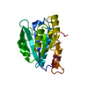
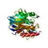
| Title | Crystal structure of the VIM-2 acquired metallo-beta-Lactamase in Complex with compound 10 (JMV-7210) | ||||||
 Components Components | Metallo-beta-lactamase VIM-2-like protein | ||||||
 Keywords Keywords |  HYDROLASE / metallo-beta-Lactamase / VIM-2 / triazole-thione / inhibitor / zinc HYDROLASE / metallo-beta-Lactamase / VIM-2 / triazole-thione / inhibitor / zinc | ||||||
| Function / homology |  Function and homology information Function and homology informationantibiotic catabolic process / beta-lactamase / hydrolase activity / metal ion bindingSimilarity search - Function | ||||||
| Biological species |   Pseudomonas aeruginosa (bacteria) Pseudomonas aeruginosa (bacteria) | ||||||
| Method | X-RAY DIFFRACTION / SYNCHROTRON / MOLECULAR REPLACEMENT / Resolution: 1.92 Å | ||||||
 Authors Authors | Tassone, G. / Benvenuti, M. / Verdirosa, F. / Sannio, F. / Mangani, S. / Docquier, J.D. / Pozzi, C. / Marcoccia, F. | ||||||
 Citation Citation | Journal: Eur.J.Med.Chem. / Year: 2021 Title: 1,2,4-Triazole-3-thione compounds with a 4-ethyl alkyl/aryl sulfide substituent are broad-spectrum metallo-beta-lactamase inhibitors with re-sensitization activity. Authors: Legru, A. / Verdirosa, F. / Hernandez, J.F. / Tassone, G. / Sannio, F. / Benvenuti, M. / Conde, P.A. / Bossis, G. / Thomas, C.A. / Crowder, M.W. / Dillenberger, M. / Becker, K. / Pozzi, C. / ...Authors: Legru, A. / Verdirosa, F. / Hernandez, J.F. / Tassone, G. / Sannio, F. / Benvenuti, M. / Conde, P.A. / Bossis, G. / Thomas, C.A. / Crowder, M.W. / Dillenberger, M. / Becker, K. / Pozzi, C. / Mangani, S. / Docquier, J.D. / Gavara, L. | ||||||
| History |
|
- Structure visualization
| Structure viewer | Molecule: MolmilJmol/JSmol |
|---|
- Downloads & links
-Download
| PDBx/mmCIF format | 7ove.cif.gz | 62.8 KB | Display | PDBx/mmCIF format |
|---|---|---|---|---|
| PDB format | pdb7ove.ent.gz | 43.7 KB | Display | PDB format |
| PDBx/mmJSON format | 7ove.json.gz | Tree view | PDBx/mmJSON format | |
| Others |  Other downloads Other downloads |
-Validation report
| Arichive directory | https://data.pdbj.org/pub/pdb/validation_reports/ov/7oveftp://data.pdbj.org/pub/pdb/validation_reports/ov/7ove | HTTPS FTP |
|---|
-Related structure data
| Related structure data |  7ovfC  7ovhC  6sp7S S: Starting model for refinement C: citing same article ( |
|---|---|
| Similar structure data |


-Links
 PDBj
PDBj


- Assembly
| Deposited unit | 
| ||||||||
|---|---|---|---|---|---|---|---|---|---|
| 1 |
| ||||||||
| Unit cell |
|
-Components
| #1: Protein | Mass: 25539.322 Da / Num. of mol.: 1 Source method: isolated from a genetically manipulated source Source: (gene. exp.) Pseudomonas aeruginosa (bacteria) / Gene: blaVIM / Plasmid: pET9 / Production host: Escherichia coli BL21(DE3) (bacteria) / References: UniProt: B8QIQ9 | ||||||||
|---|---|---|---|---|---|---|---|---|---|
| #2: Chemical |   Mass: 65.409 Da / Num. of mol.: 3 / Source method: obtained synthetically / Formula: Zn Mass: 65.409 Da / Num. of mol.: 3 / Source method: obtained synthetically / Formula: Zn#3: Chemical | Acetate  Mass: 59.044 Da / Num. of mol.: 2 / Source method: obtained synthetically / Formula: C2H3O2 Mass: 59.044 Da / Num. of mol.: 2 / Source method: obtained synthetically / Formula: C2H3O2#4: Chemical | ChemComp-UNL / | Num. of mol.: 1 / Source method: obtained synthetically / Feature type: SUBJECT OF INVESTIGATION #5: Water | ChemComp-HOH / | Water Mass: 18.015 Da / Num. of mol.: 153 / Source method: isolated from a natural source / Formula: H2O Mass: 18.015 Da / Num. of mol.: 153 / Source method: isolated from a natural source / Formula: H2OHas ligand of interest | Y | |
-Experimental details
-Experiment
| Experiment | Method: X-RAY DIFFRACTION / Number of used crystals: 1 |
|---|
- Sample preparation
| Crystal | Density Matthews: 2.34 Å3/Da / Density % sol: 47.4 % |
|---|---|
| Crystal grow | Temperature: 293 K / Method: vapor diffusion, sitting drop / pH: 6.5 Details: 0.1 M cacodilate (pH 6.5), 5 mM DTT , 0.2 M Na-acetate, 26% PEG 8000 |
-Data collection
| Diffraction | Mean temperature: 100 K / Serial crystal experiment: N |
|---|---|
| Diffraction source | Source: SYNCHROTRON / Site: Diamond  / Beamline: I04 / Wavelength: 0.979499 Å / Beamline: I04 / Wavelength: 0.979499 Å |
| Detector | Type: DECTRIS EIGER2 XE 16M / Detector: PIXEL / Date: Dec 4, 2019 |
| Radiation | Monochromator: Si(111) / Protocol: SINGLE WAVELENGTH / Monochromatic (M) / Laue (L): M / Scattering type: x-ray |
| Radiation wavelength | Wavelength: 0.979499 Å / Relative weight: 1 |
| Reflection | Resolution: 1.92→55.66 Å / Num. obs: 16503 / % possible obs: 99.4 % / Redundancy: 7.4 % / Biso Wilson estimate: 22.5 Å2 / CC1/2: 0.994 / Rmerge(I) obs: 0.124 / Rpim(I) all: 0.073 / Rrim(I) all: 0.144 / Net I/σ(I): 9.5 |
| Reflection shell | Resolution: 1.92→2.02 Å / Redundancy: 7.8 % / Rmerge(I) obs: 0.714 / Mean I/σ(I) obs: 2.5 / Num. unique obs: 2369 / CC1/2: 0.807 / Rpim(I) all: 0.41 / Rrim(I) all: 0.825 / % possible all: 100 |
- Processing
| Software |
| |||||||||||||||||||||||||||||||||||||||||||||
|---|---|---|---|---|---|---|---|---|---|---|---|---|---|---|---|---|---|---|---|---|---|---|---|---|---|---|---|---|---|---|---|---|---|---|---|---|---|---|---|---|---|---|---|---|---|---|
| Refinement | Method to determine structure: MOLECULAR REPLACEMENT Starting model: 6SP7 Resolution: 1.92→51.4 Å / Cor.coef. Fo:Fc: 0.957 / Cor.coef. Fo:Fc free: 0.929 / SU B: 4.748 / SU ML: 0.134 / Cross valid method: THROUGHOUT / σ(F): 0 / ESU R: 0.182 / ESU R Free: 0.169 / Stereochemistry target values: MAXIMUM LIKELIHOOD / Details: U VALUES : REFINED INDIVIDUALLY
| |||||||||||||||||||||||||||||||||||||||||||||
| Solvent computation | Ion probe radii: 0.8 Å / Shrinkage radii: 0.8 Å / VDW probe radii: 1.2 Å / Solvent model: MASK | |||||||||||||||||||||||||||||||||||||||||||||
| Displacement parameters | Biso max: 80.5 Å2 / Biso mean: 27.943 Å2 / Biso min: 9.13 Å2
| |||||||||||||||||||||||||||||||||||||||||||||
| Refine analyze |
| |||||||||||||||||||||||||||||||||||||||||||||
| Refinement step | Cycle: final / Resolution: 1.92→51.4 Å
| |||||||||||||||||||||||||||||||||||||||||||||
| Refine LS restraints |
| |||||||||||||||||||||||||||||||||||||||||||||
| LS refinement shell | Resolution: 1.92→1.97 Å / Rfactor Rfree error: 0 / Total num. of bins used: 20
|
