 Movie
Movie Controller

[English] 日本語
 Yorodumi
- PDB-7okv: Crystal structure of soluble EPCR after exposure to the nonionic ... -
+ Open data

- Basic information
| Entry | Database: PDB / ID: 7okv | ||||||
|---|---|---|---|---|---|---|---|
| Title | Crystal structure of soluble EPCR after exposure to the nonionic surfactant Polysorbate 20 | ||||||
 Components Components | Endothelial protein C receptor | ||||||
 Keywords Keywords | LIPID BINDING PROTEIN / MHC class-I like / phospholipid / anticoagulant / endothelial cell membrane receptor | ||||||
| Function / homology |  Function and homology information Function and homology informationnegative regulation of coagulation / Common Pathway of Fibrin Clot Formation / Cell surface interactions at the vascular wall / blood coagulation / signaling receptor activity / focal adhesion / centrosome / perinuclear region of cytoplasm / cell surface / extracellular space ...negative regulation of coagulation / Common Pathway of Fibrin Clot Formation / Cell surface interactions at the vascular wall / blood coagulation / signaling receptor activity / focal adhesion / centrosome / perinuclear region of cytoplasm / cell surface / extracellular space / extracellular exosome / extracellular region / plasma membraneSimilarity search - Function | ||||||
| Biological species |  Homo sapiens (human) Homo sapiens (human) | ||||||
| Method | X-RAY DIFFRACTION / SYNCHROTRON / MOLECULAR REPLACEMENT / Resolution: 1.85 Å | ||||||
 Authors Authors | Erausquin, E. / Dichiara, M.G. / Lopez-Sagaseta, J. | ||||||
| Funding support |  Spain, 1items Spain, 1items
| ||||||
 Citation Citation | Journal: Sci Rep / Year: 2022 Title: Identification of a broad lipid repertoire associated to the endothelial cell protein C receptor (EPCR). Authors: Erausquin, E. / Moran-Garrido, M. / Saiz, J. / Barbas, C. / Dichiara-Rodriguez, G. / Urdiciain, A. / Lopez-Sagaseta, J. | ||||||
| History |
|
- Structure visualization
| Structure viewer | Molecule: MolmilJmol/JSmol |
|---|
- Downloads & links
-Download
| PDBx/mmCIF format | 7okv.cif.gz | 106.6 KB | Display | PDBx/mmCIF format |
|---|---|---|---|---|
| PDB format | pdb7okv.ent.gz | 67.4 KB | Display | PDB format |
| PDBx/mmJSON format | 7okv.json.gz | Tree view | PDBx/mmJSON format | |
| Others |  Other downloads Other downloads |
-Validation report
| Arichive directory | https://data.pdbj.org/pub/pdb/validation_reports/ok/7okvftp://data.pdbj.org/pub/pdb/validation_reports/ok/7okv | HTTPS FTP |
|---|
-Related structure data
| Related structure data |  7oksC 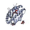 7oktC 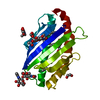 7okuC 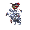 1lqvS S: Starting model for refinement C: citing same article ( |
|---|---|
| Similar structure data |
-Links
 PDBj
PDBj


- Assembly
| Deposited unit | 
| ||||||||||||
|---|---|---|---|---|---|---|---|---|---|---|---|---|---|
| 1 |
| ||||||||||||
| Unit cell |
|
-Components
-Protein / Sugars , 2 types, 4 molecules A
| #1: Protein | / Activated protein C receptor / APC receptor / Endothelial cell protein C receptor Mass: 22200.730 Da / Num. of mol.: 1 Source method: isolated from a genetically manipulated source Details: Remaining N-terminal GP motif from 3C site / Source: (gene. exp.) Homo sapiens (human) / Gene: PROCR, EPCR / Plasmid: pAcGP67A / Production host:   Spodoptera frugiperda (fall armyworm) / References: UniProt: Q9UNN8 Spodoptera frugiperda (fall armyworm) / References: UniProt: Q9UNN8 |
|---|---|
| #2: Sugar | N-Acetylglucosamine Type: D-saccharide, beta linking / Mass: 221.208 Da / Num. of mol.: 3 / Source method: obtained synthetically / Formula: C8H15NO6 Type: D-saccharide, beta linking / Mass: 221.208 Da / Num. of mol.: 3 / Source method: obtained synthetically / Formula: C8H15NO6 |
-Non-polymers , 5 types, 60 molecules 


| #3: Chemical | ChemComp-R16 / Hexadecane Mass: 226.441 Da / Num. of mol.: 1 / Source method: obtained synthetically / Formula: C16H34 / Feature type: SUBJECT OF INVESTIGATION Mass: 226.441 Da / Num. of mol.: 1 / Source method: obtained synthetically / Formula: C16H34 / Feature type: SUBJECT OF INVESTIGATION |
|---|---|
| #4: Chemical | ChemComp-HEX / Hexane Mass: 86.175 Da / Num. of mol.: 1 / Source method: obtained synthetically / Formula: C6H14 / Feature type: SUBJECT OF INVESTIGATION Mass: 86.175 Da / Num. of mol.: 1 / Source method: obtained synthetically / Formula: C6H14 / Feature type: SUBJECT OF INVESTIGATION |
| #5: Chemical | ChemComp-D10 / Decane Mass: 142.282 Da / Num. of mol.: 1 / Source method: obtained synthetically / Formula: C10H22 / Feature type: SUBJECT OF INVESTIGATION Mass: 142.282 Da / Num. of mol.: 1 / Source method: obtained synthetically / Formula: C10H22 / Feature type: SUBJECT OF INVESTIGATION |
| #6: Chemical | ChemComp-EDO / Ethylene glycol Mass: 62.068 Da / Num. of mol.: 1 / Source method: obtained synthetically / Formula: C2H6O2 Mass: 62.068 Da / Num. of mol.: 1 / Source method: obtained synthetically / Formula: C2H6O2 |
| #7: Water | ChemComp-HOH / WaterMass: 18.015 Da / Num. of mol.: 56 / Source method: isolated from a natural source / Formula: H2O |
-Details
| Has ligand of interest | Y |
|---|
-Experimental details
-Experiment
| Experiment | Method: X-RAY DIFFRACTION / Number of used crystals: 1 |
|---|
- Sample preparation
| Crystal | Density Matthews: 3.39 Å3/Da / Density % sol: 63.67 % |
|---|---|
| Crystal grow | Temperature: 295 K / Method: vapor diffusion, sitting drop / pH: 4.6 / Details: 3.5 M Sodium formate, 0.1 M Sodium acetate pH 4.6 |
-Data collection
| Diffraction | Mean temperature: 100 K / Serial crystal experiment: N |
|---|---|
| Diffraction source | Source: SYNCHROTRON / Site: ALBA / Beamline: XALOC / Wavelength: 0.97934 Å |
| Detector | Type: DECTRIS PILATUS 6M / Detector: PIXEL / Date: May 29, 2020 |
| Radiation | Monochromator: Si(111) channel-cut / Protocol: SINGLE WAVELENGTH / Monochromatic (M) / Laue (L): M / Scattering type: x-ray |
| Radiation wavelength | Wavelength: 0.97934 Å / Relative weight: 1 |
| Reflection | Resolution: 1.85→35.53 Å / Num. obs: 26239 / % possible obs: 99.8 % / Redundancy: 8.1 % / Biso Wilson estimate: 37.8 Å2 / CC1/2: 0.998 / Rmerge(I) obs: 0.098 / Rpim(I) all: 0.037 / Rrim(I) all: 0.105 / Χ2: 0.94 / Net I/σ(I): 10.6 |
| Reflection shell | Resolution: 1.85→1.89 Å / Redundancy: 8.5 % / Mean I/σ(I) obs: 1.2 / Num. unique obs: 1589 / CC1/2: 0.543 / Χ2: 0.99 / % possible all: 99.4 |
- Processing
| Software |
| |||||||||||||||||||||||||||||||||||||||||||||||||||||||||||||||||||||||||||||
|---|---|---|---|---|---|---|---|---|---|---|---|---|---|---|---|---|---|---|---|---|---|---|---|---|---|---|---|---|---|---|---|---|---|---|---|---|---|---|---|---|---|---|---|---|---|---|---|---|---|---|---|---|---|---|---|---|---|---|---|---|---|---|---|---|---|---|---|---|---|---|---|---|---|---|---|---|---|---|
| Refinement | Method to determine structure: MOLECULAR REPLACEMENT Starting model: 1LQV Resolution: 1.85→35.53 Å / SU ML: 0.2524 / Cross valid method: FREE R-VALUE / σ(F): 1.33 / Phase error: 27.6203 Stereochemistry target values: GeoStd + Monomer Library + CDL v1.2
| |||||||||||||||||||||||||||||||||||||||||||||||||||||||||||||||||||||||||||||
| Solvent computation | Shrinkage radii: 0.9 Å / VDW probe radii: 1.11 Å / Solvent model: FLAT BULK SOLVENT MODEL | |||||||||||||||||||||||||||||||||||||||||||||||||||||||||||||||||||||||||||||
| Displacement parameters | Biso mean: 51.34 Å2 | |||||||||||||||||||||||||||||||||||||||||||||||||||||||||||||||||||||||||||||
| Refinement step | Cycle: LAST / Resolution: 1.85→35.53 Å
| |||||||||||||||||||||||||||||||||||||||||||||||||||||||||||||||||||||||||||||
| Refine LS restraints |
| |||||||||||||||||||||||||||||||||||||||||||||||||||||||||||||||||||||||||||||
| LS refinement shell |
| |||||||||||||||||||||||||||||||||||||||||||||||||||||||||||||||||||||||||||||
| Refinement TLS params. | Method: refined / Origin x: 26.1429228867 Å / Origin y: -21.6680897467 Å / Origin z: 3.05524115638 Å
| |||||||||||||||||||||||||||||||||||||||||||||||||||||||||||||||||||||||||||||
| Refinement TLS group | Selection details: all |
