 Movie
Movie Controller

[English] 日本語
 Yorodumi
- PDB-7oiw: Structure of S. aureus Rel catalytic domains in complex with pppGpp -
+ Open data

- Basic information
| Entry | Database: PDB / ID: 7oiw | |||||||||
|---|---|---|---|---|---|---|---|---|---|---|
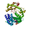
| Title | Structure of S. aureus Rel catalytic domains in complex with pppGpp | |||||||||
 Components Components | GTP pyrophosphokinase | |||||||||
 Keywords Keywords |  TRANSFERASE / bifunctional GTP pyrophosphokinase / hydrolase / alarmone synthetase / hydrolase / ppGpp TRANSFERASE / bifunctional GTP pyrophosphokinase / hydrolase / alarmone synthetase / hydrolase / ppGpp | |||||||||
| Function / homology |  Function and homology informationGTP diphosphokinase activity / guanosine tetraphosphate biosynthetic process / GTP diphosphokinase / kinase activity / GTP binding / ATP binding Function and homology informationGTP diphosphokinase activity / guanosine tetraphosphate biosynthetic process / GTP diphosphokinase / kinase activity / GTP binding / ATP bindingSimilarity search - Function | |||||||||
| Biological species |   Staphylococcus aureus (bacteria) Staphylococcus aureus (bacteria) | |||||||||
| Method | X-RAY DIFFRACTION / SYNCHROTRON / MOLECULAR REPLACEMENT / Resolution: 2.63 Å | |||||||||
 Authors Authors | Garcia-Pino, A. | |||||||||
 Citation Citation | Journal: To Be Published Title: Structure of S. aureus Rel catalytic domains in complex with pppGpp Authors: Garcia-Pino, A. | |||||||||
| History |
|
- Structure visualization
| Structure viewer | Molecule: MolmilJmol/JSmol |
|---|
- Downloads & links
-Download
| PDBx/mmCIF format | 7oiw.cif.gz | 287.6 KB | Display | PDBx/mmCIF format |
|---|---|---|---|---|
| PDB format | pdb7oiw.ent.gz | 229.1 KB | Display | PDB format |
| PDBx/mmJSON format | 7oiw.json.gz | Tree view | PDBx/mmJSON format | |
| Others |  Other downloads Other downloads |
-Validation report
| Arichive directory | https://data.pdbj.org/pub/pdb/validation_reports/oi/7oiwftp://data.pdbj.org/pub/pdb/validation_reports/oi/7oiw | HTTPS FTP |
|---|
-Related structure data
| Related structure data | 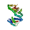 6s2uS S: Starting model for refinement |
|---|---|
| Similar structure data |
-Links
 PDBj
PDBj


- Assembly
| Deposited unit | 
| ||||||||||
|---|---|---|---|---|---|---|---|---|---|---|---|
| 1 | 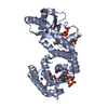
| ||||||||||
| 2 | 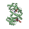
| ||||||||||
| Unit cell |
|
-Components
-Protein , 1 types, 2 molecules AB
| #1: Protein | Mass: 51057.574 Da / Num. of mol.: 2 Source method: isolated from a genetically manipulated source Details: the missing residues are not visible in the electron density Source: (gene. exp.) Staphylococcus aureus (strain Mu50 / ATCC 700699) (bacteria)Strain: Mu50 / ATCC 700699 / Gene: relA, SAV1634 / Production host: Escherichia coli (E. coli) / References: UniProt: Q931Q4, GTP diphosphokinase |
|---|
-Non-polymers , 7 types, 78 molecules 


| #2: Chemical | ChemComp-0O2 /  Mass: 683.140 Da / Num. of mol.: 4 / Source method: obtained synthetically / Formula: C10H18N5O20P5 / Feature type: SUBJECT OF INVESTIGATION Mass: 683.140 Da / Num. of mol.: 4 / Source method: obtained synthetically / Formula: C10H18N5O20P5 / Feature type: SUBJECT OF INVESTIGATION#3: Chemical | ChemComp-VF5 / [[( |  Mass: 585.145 Da / Num. of mol.: 1 / Source method: obtained synthetically / Formula: C10H15N5O16P4 Mass: 585.145 Da / Num. of mol.: 1 / Source method: obtained synthetically / Formula: C10H15N5O16P4#4: Chemical | ChemComp-GOL / | Glycerol Mass: 92.094 Da / Num. of mol.: 1 / Source method: obtained synthetically / Formula: C3H8O3 Mass: 92.094 Da / Num. of mol.: 1 / Source method: obtained synthetically / Formula: C3H8O3#5: Chemical |  Mass: 54.938 Da / Num. of mol.: 2 / Source method: obtained synthetically / Formula: Mn Mass: 54.938 Da / Num. of mol.: 2 / Source method: obtained synthetically / Formula: Mn#6: Chemical |  Mass: 24.305 Da / Num. of mol.: 3 / Source method: obtained synthetically / Formula: Mg Mass: 24.305 Da / Num. of mol.: 3 / Source method: obtained synthetically / Formula: Mg#7: Chemical | ChemComp-IOD / Iodide Mass: 126.904 Da / Num. of mol.: 12 / Source method: obtained synthetically / Formula: I Mass: 126.904 Da / Num. of mol.: 12 / Source method: obtained synthetically / Formula: I#8: Water | ChemComp-HOH / | WaterMass: 18.015 Da / Num. of mol.: 55 / Source method: isolated from a natural source / Formula: H2O |
|---|
-Details
| Has ligand of interest | Y |
|---|
-Experimental details
-Experiment
| Experiment | Method: X-RAY DIFFRACTION / Number of used crystals: 1 |
|---|
- Sample preparation
| Crystal | Density Matthews: 3.48 Å3/Da / Density % sol: 64.64 % |
|---|---|
| Crystal grow | Temperature: 293.16 K / Method: vapor diffusion, sitting drop Details: 6% w/vPEG 3350 0.15 M Sodium chloride 0.4 M Potassium iodide pH 8.5 |
-Data collection
| Diffraction | Mean temperature: 100 K / Serial crystal experiment: N |
|---|---|
| Diffraction source | Source: SYNCHROTRON / Site: SOLEIL  / Beamline: PROXIMA 2 / Wavelength: 0.9801 Å / Beamline: PROXIMA 2 / Wavelength: 0.9801 Å |
| Detector | Type: DECTRIS PILATUS 12M / Detector: PIXEL / Date: Mar 22, 2019 |
| Radiation | Protocol: SINGLE WAVELENGTH / Monochromatic (M) / Laue (L): M / Scattering type: x-ray |
| Radiation wavelength | Wavelength: 0.9801 Å / Relative weight: 1 |
| Reflection | Resolution: 2.63→42.25 Å / Num. obs: 31024 / % possible obs: 99.4 % / Redundancy: 7.3 % / Biso Wilson estimate: 59.19 Å2 / CC1/2: 0.993 / Rmerge(I) obs: 0.18 / Rpim(I) all: 0.07 / Net I/σ(I): 8.2 |
| Reflection shell | Resolution: 2.63→2.72 Å / Rmerge(I) obs: 1.21 / Mean I/σ(I) obs: 1.2 / Num. unique obs: 2954 / CC1/2: 0.667 / Rpim(I) all: 0.47 |
- Processing
| Software |
| |||||||||||||||||||||||||||||||||||||||||||||||||||||||||||||||||||||||||||||||||||||||||||||||||||||||||||||||||||||||||||||||||||||||||||||||||||||||||||||||||||||||||||||||||||||||||||||||||||||||||||||||||||||||||||||||||||||||||||||||||||||||||||||||||||||||||||||||||||
|---|---|---|---|---|---|---|---|---|---|---|---|---|---|---|---|---|---|---|---|---|---|---|---|---|---|---|---|---|---|---|---|---|---|---|---|---|---|---|---|---|---|---|---|---|---|---|---|---|---|---|---|---|---|---|---|---|---|---|---|---|---|---|---|---|---|---|---|---|---|---|---|---|---|---|---|---|---|---|---|---|---|---|---|---|---|---|---|---|---|---|---|---|---|---|---|---|---|---|---|---|---|---|---|---|---|---|---|---|---|---|---|---|---|---|---|---|---|---|---|---|---|---|---|---|---|---|---|---|---|---|---|---|---|---|---|---|---|---|---|---|---|---|---|---|---|---|---|---|---|---|---|---|---|---|---|---|---|---|---|---|---|---|---|---|---|---|---|---|---|---|---|---|---|---|---|---|---|---|---|---|---|---|---|---|---|---|---|---|---|---|---|---|---|---|---|---|---|---|---|---|---|---|---|---|---|---|---|---|---|---|---|---|---|---|---|---|---|---|---|---|---|---|---|---|---|---|---|---|---|---|---|---|---|---|---|---|---|---|---|---|---|---|---|---|---|---|---|---|---|---|---|---|---|---|---|---|---|---|---|---|---|---|---|---|---|---|---|---|---|---|---|---|---|---|---|---|
| Refinement | Method to determine structure: MOLECULAR REPLACEMENT Starting model: 6S2U Resolution: 2.63→42.25 Å / SU ML: 0.38 / Cross valid method: FREE R-VALUE / σ(F): 1.38 / Phase error: 26.24 / Stereochemistry target values: ML
| |||||||||||||||||||||||||||||||||||||||||||||||||||||||||||||||||||||||||||||||||||||||||||||||||||||||||||||||||||||||||||||||||||||||||||||||||||||||||||||||||||||||||||||||||||||||||||||||||||||||||||||||||||||||||||||||||||||||||||||||||||||||||||||||||||||||||||||||||||
| Solvent computation | Shrinkage radii: 0.9 Å / VDW probe radii: 1.11 Å / Solvent model: FLAT BULK SOLVENT MODEL | |||||||||||||||||||||||||||||||||||||||||||||||||||||||||||||||||||||||||||||||||||||||||||||||||||||||||||||||||||||||||||||||||||||||||||||||||||||||||||||||||||||||||||||||||||||||||||||||||||||||||||||||||||||||||||||||||||||||||||||||||||||||||||||||||||||||||||||||||||
| Refinement step | Cycle: LAST / Resolution: 2.63→42.25 Å
| |||||||||||||||||||||||||||||||||||||||||||||||||||||||||||||||||||||||||||||||||||||||||||||||||||||||||||||||||||||||||||||||||||||||||||||||||||||||||||||||||||||||||||||||||||||||||||||||||||||||||||||||||||||||||||||||||||||||||||||||||||||||||||||||||||||||||||||||||||
| Refine LS restraints |
| |||||||||||||||||||||||||||||||||||||||||||||||||||||||||||||||||||||||||||||||||||||||||||||||||||||||||||||||||||||||||||||||||||||||||||||||||||||||||||||||||||||||||||||||||||||||||||||||||||||||||||||||||||||||||||||||||||||||||||||||||||||||||||||||||||||||||||||||||||
| LS refinement shell |
| |||||||||||||||||||||||||||||||||||||||||||||||||||||||||||||||||||||||||||||||||||||||||||||||||||||||||||||||||||||||||||||||||||||||||||||||||||||||||||||||||||||||||||||||||||||||||||||||||||||||||||||||||||||||||||||||||||||||||||||||||||||||||||||||||||||||||||||||||||
| Refinement TLS params. | Method: refined / Refine-ID: X-RAY DIFFRACTION
| |||||||||||||||||||||||||||||||||||||||||||||||||||||||||||||||||||||||||||||||||||||||||||||||||||||||||||||||||||||||||||||||||||||||||||||||||||||||||||||||||||||||||||||||||||||||||||||||||||||||||||||||||||||||||||||||||||||||||||||||||||||||||||||||||||||||||||||||||||
| Refinement TLS group |
|
