 Movie
Movie Controller

[English] 日本語
 Yorodumi
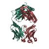
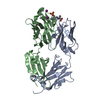
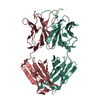
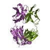
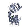
- PDB-6s2u: Structure of the catalytic domain of T. thermophilus Rel in compl... -
+ Open data

- Basic information
| Entry | Database: PDB / ID: 6s2u | ||||||
|---|---|---|---|---|---|---|---|
| Title | Structure of the catalytic domain of T. thermophilus Rel in complex with AMP and ppGpp | ||||||
 Components Components | (P)ppGpp synthetase I, SpoT/RelA | ||||||
 Keywords Keywords |  TRANSFERASE / ppGpp synthetase / ppGpp hydrolase / ppGpp / translation / stringent response TRANSFERASE / ppGpp synthetase / ppGpp hydrolase / ppGpp / translation / stringent response | ||||||
| Function / homology |  Function and homology information Function and homology informationguanosine tetraphosphate metabolic process / GTP diphosphokinase activity / GTP diphosphokinaseSimilarity search - Function | ||||||
| Biological species |   Thermus thermophilus (bacteria) Thermus thermophilus (bacteria) | ||||||
| Method | X-RAY DIFFRACTION / SYNCHROTRON / MOLECULAR REPLACEMENT / Resolution: 2.95 Å | ||||||
 Authors Authors | Garcia-Pino, A. | ||||||
 Citation Citation | Journal: Nat.Chem.Biol. / Year: 2020 Title: A nucleotide-switch mechanism mediates opposing catalytic activities of Rel enzymes. Authors: Tamman, H. / Van Nerom, K. / Takada, H. / Vandenberk, N. / Scholl, D. / Polikanov, Y. / Hofkens, J. / Talavera, A. / Hauryliuk, V. / Hendrix, J. / Garcia-Pino, A. | ||||||
| History |
|
- Structure visualization
| Structure viewer | Molecule: MolmilJmol/JSmol |
|---|
- Downloads & links
-Download
| PDBx/mmCIF format | 6s2u.cif.gz | 153.3 KB | Display | PDBx/mmCIF format |
|---|---|---|---|---|
| PDB format | pdb6s2u.ent.gz | 120.3 KB | Display | PDB format |
| PDBx/mmJSON format | 6s2u.json.gz | Tree view | PDBx/mmJSON format | |
| Others |  Other downloads Other downloads |
-Validation report
| Arichive directory | https://data.pdbj.org/pub/pdb/validation_reports/s2/6s2uftp://data.pdbj.org/pub/pdb/validation_reports/s2/6s2u | HTTPS FTP |
|---|
-Related structure data
| Related structure data |  6s2tC  6s2vC 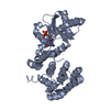 1vj7S S: Starting model for refinement C: citing same article ( |
|---|---|
| Similar structure data |

-Links
 PDBj
PDBj


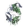
- Assembly
| Deposited unit | 
| ||||||||
|---|---|---|---|---|---|---|---|---|---|
| 1 |
| ||||||||
| Unit cell |
| ||||||||
| Components on special symmetry positions |
|
-Components
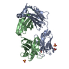
-Protein , 1 types, 1 molecules A
| #1: Protein | ( Mass: 40705.883 Da / Num. of mol.: 1 Source method: isolated from a genetically manipulated source Source: (gene. exp.) Thermus thermophilus (bacteria) / Gene: Ththe16_1734 / Production host: Escherichia coli (E. coli)References: UniProt: F6DES6, UniProt: Q5SHL3*PLUS, GTP diphosphokinase |
|---|
-Non-polymers , 6 types, 27 molecules 


| #2: Chemical | ChemComp-MN /  Mass: 54.938 Da / Num. of mol.: 1 / Source method: obtained synthetically / Formula: Mn Mass: 54.938 Da / Num. of mol.: 1 / Source method: obtained synthetically / Formula: Mn | ||
|---|---|---|---|
| #3: Chemical | ChemComp-AMP / Adenosine monophosphate Mass: 347.221 Da / Num. of mol.: 1 / Source method: obtained synthetically / Formula: C10H14N5O7P / Comment: AMP*YM Mass: 347.221 Da / Num. of mol.: 1 / Source method: obtained synthetically / Formula: C10H14N5O7P / Comment: AMP*YM | ||
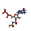
| #4: Chemical | ChemComp-GN3 / [[[( Mass: 602.176 Da / Num. of mol.: 1 / Source method: obtained synthetically / Formula: C10H18N6O16P4 Mass: 602.176 Da / Num. of mol.: 1 / Source method: obtained synthetically / Formula: C10H18N6O16P4 | ||
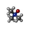
| #5: Chemical | ChemComp-TMO / Trimethylamine N-oxide Mass: 75.110 Da / Num. of mol.: 1 / Source method: obtained synthetically / Formula: C3H9NO Mass: 75.110 Da / Num. of mol.: 1 / Source method: obtained synthetically / Formula: C3H9NO | ||
| #6: Chemical |  Mass: 24.305 Da / Num. of mol.: 2 / Source method: obtained synthetically / Formula: Mg Mass: 24.305 Da / Num. of mol.: 2 / Source method: obtained synthetically / Formula: Mg#7: Water | ChemComp-HOH / | WaterMass: 18.015 Da / Num. of mol.: 21 / Source method: isolated from a natural source / Formula: H2O |
-Details
| Has ligand of interest | N |
|---|
-Experimental details
-Experiment
| Experiment | Method: X-RAY DIFFRACTION / Number of used crystals: 1 |
|---|
- Sample preparation
| Crystal | Density Matthews: 3 Å3/Da / Density % sol: 58.99 % |
|---|---|
| Crystal grow | Temperature: 293 K / Method: vapor diffusion, sitting drop / pH: 8 Details: 0.002 M Zinc chloride 0.1 M Tris 8.0 20 % w/v PEG 6000 |
-Data collection
| Diffraction | Mean temperature: 100 K / Serial crystal experiment: N |
|---|---|
| Diffraction source | Source: SYNCHROTRON / Site: SOLEIL  / Beamline: PROXIMA 2 / Wavelength: 0.9801 Å / Beamline: PROXIMA 2 / Wavelength: 0.9801 Å |
| Detector | Type: DECTRIS EIGER X 9M / Detector: PIXEL / Date: Jul 1, 2016 |
| Radiation | Protocol: SINGLE WAVELENGTH / Monochromatic (M) / Laue (L): M / Scattering type: x-ray |
| Radiation wavelength | Wavelength: 0.9801 Å / Relative weight: 1 |
| Reflection | Resolution: 2.95→56.36 Å / Num. obs: 6572 / % possible obs: 91.9 % / Redundancy: 10.3 % / Biso Wilson estimate: 102.64 Å2 / CC1/2: 0.996 / Rmerge(I) obs: 0.12 / Rpim(I) all: 0.04 / Net I/σ(I): 13.8 |
| Reflection shell | Resolution: 2.95→3.02 Å / Rmerge(I) obs: 1.1 / Num. unique obs: 2399 / CC1/2: 0.868 / Rpim(I) all: 0.36 |
- Processing
| Software |
| ||||||||||||||||||||||||||||||||||||||||||||||||||||||||||||||||||||||||||||||||||||||||||||||||||||||||||||||||||
|---|---|---|---|---|---|---|---|---|---|---|---|---|---|---|---|---|---|---|---|---|---|---|---|---|---|---|---|---|---|---|---|---|---|---|---|---|---|---|---|---|---|---|---|---|---|---|---|---|---|---|---|---|---|---|---|---|---|---|---|---|---|---|---|---|---|---|---|---|---|---|---|---|---|---|---|---|---|---|---|---|---|---|---|---|---|---|---|---|---|---|---|---|---|---|---|---|---|---|---|---|---|---|---|---|---|---|---|---|---|---|---|---|---|---|---|
| Refinement | Method to determine structure: MOLECULAR REPLACEMENT Starting model: 1vj7 Resolution: 2.95→56.36 Å / Cor.coef. Fo:Fc: 0.909 / Cor.coef. Fo:Fc free: 0.879 / Cross valid method: THROUGHOUT / σ(F): 0 / SU Rfree Blow DPI: 0.528
| ||||||||||||||||||||||||||||||||||||||||||||||||||||||||||||||||||||||||||||||||||||||||||||||||||||||||||||||||||
| Displacement parameters | Biso mean: 101.56 Å2
| ||||||||||||||||||||||||||||||||||||||||||||||||||||||||||||||||||||||||||||||||||||||||||||||||||||||||||||||||||
| Refine analyze | Luzzati coordinate error obs: 0.46 Å | ||||||||||||||||||||||||||||||||||||||||||||||||||||||||||||||||||||||||||||||||||||||||||||||||||||||||||||||||||
| Refinement step | Cycle: LAST / Resolution: 2.95→56.36 Å
| ||||||||||||||||||||||||||||||||||||||||||||||||||||||||||||||||||||||||||||||||||||||||||||||||||||||||||||||||||
| Refine LS restraints |
| ||||||||||||||||||||||||||||||||||||||||||||||||||||||||||||||||||||||||||||||||||||||||||||||||||||||||||||||||||
| LS refinement shell | Resolution: 2.95→3.12 Å / Total num. of bins used: 16
| ||||||||||||||||||||||||||||||||||||||||||||||||||||||||||||||||||||||||||||||||||||||||||||||||||||||||||||||||||
| Refinement TLS params. | Method: refined / Origin x: 97.0037 Å / Origin y: -62.0508 Å / Origin z: 97.8665 Å
| ||||||||||||||||||||||||||||||||||||||||||||||||||||||||||||||||||||||||||||||||||||||||||||||||||||||||||||||||||
| Refinement TLS group | Selection details: { A|* } |
