 Movie
Movie Controller

[English] 日本語
 Yorodumi
- PDB-7ofx: Crystal structure of a GH31 family sulfoquinovosidase mutant D455... -
+ Open data

- Basic information
| Entry | Database: PDB / ID: 7ofx | ||||||
|---|---|---|---|---|---|---|---|
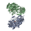
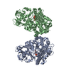
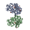
| Title | Crystal structure of a GH31 family sulfoquinovosidase mutant D455N from Agrobacterium tumefaciens in complex with sulfoquinovosyl glycerol (SQGro) | ||||||
 Components Components | Alpha-glucosidase yihQ | ||||||
 Keywords Keywords |  HYDROLASE / sulfoquinovose / SQGro / sulfoquinovosyl glycerol / SQDG / sulfoglycolysis / sulfo-EMP HYDROLASE / sulfoquinovose / SQGro / sulfoquinovosyl glycerol / SQDG / sulfoglycolysis / sulfo-EMP | ||||||
| Function / homology |  Function and homology informationalpha-glucosidase / maltose alpha-glucosidase activity / carbohydrate binding Function and homology informationalpha-glucosidase / maltose alpha-glucosidase activity / carbohydrate bindingSimilarity search - Function | ||||||
| Biological species |  Rhizobium radiobacter (bacteria) Rhizobium radiobacter (bacteria) | ||||||
| Method | X-RAY DIFFRACTION / SYNCHROTRON / MOLECULAR REPLACEMENT / Resolution: 2.15 Å | ||||||
 Authors Authors | Sharma, M. / Davies, G.J. | ||||||
| Funding support |  United Kingdom, 1items United Kingdom, 1items
| ||||||
 Citation Citation | Journal: Proc.Natl.Acad.Sci.USA / Year: 2022 Title: Oxidative desulfurization pathway for complete catabolism of sulfoquinovose by bacteria. Authors: Sharma, M. / Lingford, J.P. / Petricevic, M. / Snow, A.J.D. / Zhang, Y. / Jarva, M.A. / Mui, J.W. / Scott, N.E. / Saunders, E.C. / Mao, R. / Epa, R. / da Silva, B.M. / Pires, D.E.V. / ...Authors: Sharma, M. / Lingford, J.P. / Petricevic, M. / Snow, A.J.D. / Zhang, Y. / Jarva, M.A. / Mui, J.W. / Scott, N.E. / Saunders, E.C. / Mao, R. / Epa, R. / da Silva, B.M. / Pires, D.E.V. / Ascher, D.B. / McConville, M.J. / Davies, G.J. / Williams, S.J. / Goddard-Borger, E.D. | ||||||
| History |
|
- Structure visualization
| Structure viewer | Molecule: MolmilJmol/JSmol |
|---|
- Downloads & links
-Download
| PDBx/mmCIF format | 7ofx.cif.gz | 509.1 KB | Display | PDBx/mmCIF format |
|---|---|---|---|---|
| PDB format | pdb7ofx.ent.gz | 416.1 KB | Display | PDB format |
| PDBx/mmJSON format | 7ofx.json.gz | Tree view | PDBx/mmJSON format | |
| Others |  Other downloads Other downloads |
-Validation report
| Arichive directory | https://data.pdbj.org/pub/pdb/validation_reports/of/7ofxftp://data.pdbj.org/pub/pdb/validation_reports/of/7ofx | HTTPS FTP |
|---|
-Related structure data
| Related structure data |  7bbyC  7bbzC  7bc0C  7bc1C  7nbzC  7ofyC  7oh2C  7olfC  5ohtS S: Starting model for refinement C: citing same article ( |
|---|---|
| Similar structure data |


-Links
 PDBj
PDBj


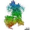
- Assembly
| Deposited unit | 
| ||||||||||||||||||||||||||||||||||||||||||||||||||||||||||||||||||||||||||||||||||||||||||||||||||||||||||||||||||||||||||||||||||||||||||||||||||||||||||||||||||||||||||||||||
|---|---|---|---|---|---|---|---|---|---|---|---|---|---|---|---|---|---|---|---|---|---|---|---|---|---|---|---|---|---|---|---|---|---|---|---|---|---|---|---|---|---|---|---|---|---|---|---|---|---|---|---|---|---|---|---|---|---|---|---|---|---|---|---|---|---|---|---|---|---|---|---|---|---|---|---|---|---|---|---|---|---|---|---|---|---|---|---|---|---|---|---|---|---|---|---|---|---|---|---|---|---|---|---|---|---|---|---|---|---|---|---|---|---|---|---|---|---|---|---|---|---|---|---|---|---|---|---|---|---|---|---|---|---|---|---|---|---|---|---|---|---|---|---|---|---|---|---|---|---|---|---|---|---|---|---|---|---|---|---|---|---|---|---|---|---|---|---|---|---|---|---|---|---|---|---|---|---|
| 1 | 
| ||||||||||||||||||||||||||||||||||||||||||||||||||||||||||||||||||||||||||||||||||||||||||||||||||||||||||||||||||||||||||||||||||||||||||||||||||||||||||||||||||||||||||||||||
| 2 | 
| ||||||||||||||||||||||||||||||||||||||||||||||||||||||||||||||||||||||||||||||||||||||||||||||||||||||||||||||||||||||||||||||||||||||||||||||||||||||||||||||||||||||||||||||||
| Unit cell |
| ||||||||||||||||||||||||||||||||||||||||||||||||||||||||||||||||||||||||||||||||||||||||||||||||||||||||||||||||||||||||||||||||||||||||||||||||||||||||||||||||||||||||||||||||
| Noncrystallographic symmetry (NCS) | NCS domain:
NCS domain segments: Component-ID: 0 / Refine code: 0
NCS ensembles :
|
-Components
| #1: Protein | Mass: 75231.438 Da / Num. of mol.: 4 / Mutation: D455N Source method: isolated from a genetically manipulated source Source: (gene. exp.) Rhizobium radiobacter (bacteria) / Gene: SY94_3281 / Production host: Escherichia coli BL21(DE3) (bacteria) / References: UniProt: A0A083ZKV2, alpha-glucosidase#2: Chemical | ChemComp-VCW / [(   Mass: 318.298 Da / Num. of mol.: 4 / Source method: obtained synthetically / Formula: C9H18O10S / Feature type: SUBJECT OF INVESTIGATION Mass: 318.298 Da / Num. of mol.: 4 / Source method: obtained synthetically / Formula: C9H18O10S / Feature type: SUBJECT OF INVESTIGATION#3: Water | ChemComp-HOH / | Water Mass: 18.015 Da / Num. of mol.: 439 / Source method: isolated from a natural source / Formula: H2O Mass: 18.015 Da / Num. of mol.: 439 / Source method: isolated from a natural source / Formula: H2OHas ligand of interest | Y | |
|---|
-Experimental details
-Experiment
| Experiment | Method: X-RAY DIFFRACTION / Number of used crystals: 1 |
|---|
- Sample preparation
| Crystal | Density Matthews: 2.49 Å3/Da / Density % sol: 50.73 % |
|---|---|
| Crystal grow | Temperature: 293 K / Method: vapor diffusion Details: 24% PEG 3350 w/v, 0.2 M KSCN, 0.1 M Bis-Tris propane pH 6.5 |
-Data collection
| Diffraction | Mean temperature: 100 K / Serial crystal experiment: N | |||||||||||||||||||||||||||
|---|---|---|---|---|---|---|---|---|---|---|---|---|---|---|---|---|---|---|---|---|---|---|---|---|---|---|---|---|
| Diffraction source | Source: SYNCHROTRON / Site: Diamond / Beamline: I03 / Wavelength: 0.9762 Å | |||||||||||||||||||||||||||
| Detector | Type: DECTRIS EIGER2 XE 16M / Detector: PIXEL / Date: Jul 28, 2019 | |||||||||||||||||||||||||||
| Radiation | Protocol: SINGLE WAVELENGTH / Monochromatic (M) / Laue (L): M / Scattering type: x-ray | |||||||||||||||||||||||||||
| Radiation wavelength | Wavelength: 0.9762 Å / Relative weight: 1 | |||||||||||||||||||||||||||
| Reflection | Resolution: 2.15→61.51 Å / Num. obs: 153301 / % possible obs: 97.4 % / Redundancy: 2.9 % / CC1/2: 0.993 / Rmerge(I) obs: 0.09 / Rpim(I) all: 0.061 / Rrim(I) all: 0.109 / Net I/σ(I): 8.1 / Num. measured all: 442708 / Scaling rejects: 162 | |||||||||||||||||||||||||||
| Reflection shell | Diffraction-ID: 1 / Redundancy: 2.9 %
|
- Processing
| Software |
| |||||||||||||||||||||||||||||||||||||||||||||||||||||||||||||||||
|---|---|---|---|---|---|---|---|---|---|---|---|---|---|---|---|---|---|---|---|---|---|---|---|---|---|---|---|---|---|---|---|---|---|---|---|---|---|---|---|---|---|---|---|---|---|---|---|---|---|---|---|---|---|---|---|---|---|---|---|---|---|---|---|---|---|---|
| Refinement | Method to determine structure: MOLECULAR REPLACEMENT Starting model: 5OHT Resolution: 2.15→61.01 Å / Cor.coef. Fo:Fc: 0.952 / Cor.coef. Fo:Fc free: 0.94 / SU B: 6.942 / SU ML: 0.167 / Cross valid method: THROUGHOUT / σ(F): 0 / ESU R: 0.249 / ESU R Free: 0.187 / Stereochemistry target values: MAXIMUM LIKELIHOOD Details: HYDROGENS HAVE BEEN ADDED IN THE RIDING POSITIONS U VALUES : REFINED INDIVIDUALLY
| |||||||||||||||||||||||||||||||||||||||||||||||||||||||||||||||||
| Solvent computation | Ion probe radii: 0.8 Å / Shrinkage radii: 0.8 Å / VDW probe radii: 1.2 Å / Solvent model: MASK | |||||||||||||||||||||||||||||||||||||||||||||||||||||||||||||||||
| Displacement parameters | Biso max: 111.95 Å2 / Biso mean: 28.618 Å2 / Biso min: 2.84 Å2
| |||||||||||||||||||||||||||||||||||||||||||||||||||||||||||||||||
| Refinement step | Cycle: final / Resolution: 2.15→61.01 Å
| |||||||||||||||||||||||||||||||||||||||||||||||||||||||||||||||||
| Refine LS restraints |
| |||||||||||||||||||||||||||||||||||||||||||||||||||||||||||||||||
| Refine LS restraints NCS | Refine-ID: X-RAY DIFFRACTION / Type: interatomic distance / Weight position: 0.05
| |||||||||||||||||||||||||||||||||||||||||||||||||||||||||||||||||
| LS refinement shell | Resolution: 2.15→2.206 Å / Rfactor Rfree error: 0 / Total num. of bins used: 20
|
