Movie
Movie Controller

[English] 日本語
 Yorodumi
- PDB-7o7f: Structural basis of the activation of the CC chemokine receptor 5... -
+ Open data

- Basic information
| Entry | Database: PDB / ID: 7o7f | |||||||||||||||
|---|---|---|---|---|---|---|---|---|---|---|---|---|---|---|---|---|
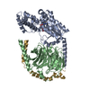
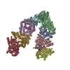
| Title | Structural basis of the activation of the CC chemokine receptor 5 by a chemokine agonist | |||||||||||||||
 Components Components |
| |||||||||||||||
 Keywords Keywords |  SIGNALING PROTEIN / G protein-coupled receptor (GPCR) / CCR5 / CCL5/RANTES / HIV entry / AIDS / membrane protein structure / MEMBRANE PROTEIN SIGNALING PROTEIN / G protein-coupled receptor (GPCR) / CCR5 / CCL5/RANTES / HIV entry / AIDS / membrane protein structure / MEMBRANE PROTEIN | |||||||||||||||
| Function / homology |  Function and homology information Function and homology informationregulation of chronic inflammatory response / CCR4 chemokine receptor binding / chemokine receptor antagonist activity / activation of phospholipase D activity / chemokine (C-C motif) ligand 5 binding / positive regulation of cellular biosynthetic process / CCR1 chemokine receptor binding / positive regulation of natural killer cell chemotaxis / negative regulation of macrophage apoptotic process / chemokine receptor binding ...regulation of chronic inflammatory response / CCR4 chemokine receptor binding / chemokine receptor antagonist activity / activation of phospholipase D activity / chemokine (C-C motif) ligand 5 binding / positive regulation of cellular biosynthetic process / CCR1 chemokine receptor binding / positive regulation of natural killer cell chemotaxis / negative regulation of macrophage apoptotic process / chemokine receptor binding / positive regulation of cell-cell adhesion mediated by integrin / signaling / Olfactory Signaling Pathway / positive regulation of activation of Janus kinase activity / Sensory perception of sweet, bitter, and umami (glutamate) taste / Synthesis, secretion, and inactivation of Glucagon-like Peptide-1 (GLP-1) / chemokine receptor activity / receptor signaling protein tyrosine kinase activator activity / positive regulation of homotypic cell-cell adhesion / CCR5 chemokine receptor binding / positive regulation of T cell chemotaxis / eye photoreceptor cell development / negative regulation of G protein-coupled receptor signaling pathway / C-C chemokine receptor activity / CCR chemokine receptor binding / lymphocyte chemotaxis / neutrophil activation / Inactivation, recovery and regulation of the phototransduction cascade / C-C chemokine binding / Activation of the phototransduction cascade / phosphatidylinositol phospholipase C activity / negative regulation of T cell apoptotic process / positive regulation of T cell apoptotic process / positive regulation of calcium ion transport / eosinophil chemotaxis / response to cholesterol / positive regulation of innate immune response / cellular response to fibroblast growth factor stimulus / chemokine-mediated signaling pathway / positive regulation of monocyte chemotaxis / Activation of G protein gated Potassium channels / G-protein activation / G beta:gamma signalling through PI3Kgamma / Prostacyclin signalling through prostacyclin receptor / G beta:gamma signalling through PLC beta / ADP signalling through P2Y purinoceptor 1 / Thromboxane signalling through TP receptor / Presynaptic function of Kainate receptors / G beta:gamma signalling through CDC42 / Inhibition of voltage gated Ca2+ channels via Gbeta/gamma subunits / Glucagon-type ligand receptors / Chemokine receptors bind chemokines / Adrenaline,noradrenaline inhibits insulin secretion / G alpha (12/13) signalling events / G beta:gamma signalling through BTK / ADP signalling through P2Y purinoceptor 12 / release of sequestered calcium ion into cytosol by sarcoplasmic reticulum / chemokine activity / Cooperation of PDCL (PhLP1) and TRiC/CCT in G-protein beta folding / dendritic cell chemotaxis / Thrombin signalling through proteinase activated receptors (PARs) / Ca2+ pathway / G alpha (z) signalling events / Extra-nuclear estrogen signaling / regulation of T cell activation / G alpha (s) signalling events / G alpha (q) signalling events / G alpha (i) signalling events / Glucagon-like Peptide-1 (GLP1) regulates insulin secretion / leukocyte cell-cell adhesion / Vasopressin regulates renal water homeostasis via Aquaporins / positive regulation of macrophage chemotaxis / negative regulation of viral genome replication / phospholipase activator activity / macrophage chemotaxis / positive regulation of smooth muscle cell migration / exocytosis / chemoattractant activity / Interleukin-10 signaling / monocyte chemotaxis / positive regulation of translational initiation / regulation of insulin secretion / phototransduction / Adenylate cyclase inhibitory pathway / positive regulation of cell adhesion / positive regulation of protein localization to cell cortex / regulation of cAMP-mediated signaling / negative regulation by host of viral transcription / positive regulation of T cell migration / D2 dopamine receptor binding / G protein-coupled serotonin receptor binding / positive regulation of viral genome replication / Binding and entry of HIV virion / cellular response to interleukin-1 / cellular defense response / coreceptor activity / regulation of mitotic spindle organization / cellular response to forskolin / positive regulation of phosphorylation / positive regulation of tyrosine phosphorylation of STAT proteinSimilarity search - Function | |||||||||||||||
| Biological species |  Homo sapiens (human) Homo sapiens (human) Mus musculus (house mouse)Bos taurus (cattle) Mus musculus (house mouse)Bos taurus (cattle) | |||||||||||||||
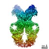
| Method | ELECTRON MICROSCOPY / single particle reconstruction / cryo EM / Resolution: 3.15 Å | |||||||||||||||
 Authors Authors | Isaikina, P. / Tsai, C.-J. / Dietz, N.B. / Pamula, F. / Goldie, K.N. / Schertler, G.F.X. / Maier, T. / Stahlberg, H. / Deupi, X. / Grzesiek, S. | |||||||||||||||
| Funding support |  Switzerland, European Union, 4items Switzerland, European Union, 4items
| |||||||||||||||
 Citation Citation | Journal: Sci Adv / Year: 2021 Title: Structural basis of the activation of the CC chemokine receptor 5 by a chemokine agonist. Authors: Polina Isaikina / Ching-Ju Tsai / Nikolaus Dietz / Filip Pamula / Anne Grahl / Kenneth N Goldie / Ramon Guixà-González / Camila Branco / Marianne Paolini-Bertrand / Nicolas Calo / Fabrice ...Authors: Polina Isaikina / Ching-Ju Tsai / Nikolaus Dietz / Filip Pamula / Anne Grahl / Kenneth N Goldie / Ramon Guixà-González / Camila Branco / Marianne Paolini-Bertrand / Nicolas Calo / Fabrice Cerini / Gebhard F X Schertler / Oliver Hartley / Henning Stahlberg / Timm Maier / Xavier Deupi / Stephan Grzesiek /  Abstract: The human CC chemokine receptor 5 (CCR5) is a G protein-coupled receptor (GPCR) that plays a major role in inflammation and is involved in cancer, HIV, and COVID-19. Despite its importance as a drug ...The human CC chemokine receptor 5 (CCR5) is a G protein-coupled receptor (GPCR) that plays a major role in inflammation and is involved in cancer, HIV, and COVID-19. Despite its importance as a drug target, the molecular activation mechanism of CCR5, i.e., how chemokine agonists transduce the activation signal through the receptor, is yet unknown. Here, we report the cryo-EM structure of wild-type CCR5 in an active conformation bound to the chemokine super-agonist [6P4]CCL5 and the heterotrimeric G protein. The structure provides the rationale for the sequence-activity relation of agonist and antagonist chemokines. The N terminus of agonist chemokines pushes onto specific structural motifs at the bottom of the orthosteric pocket that activate the canonical GPCR microswitch network. This activation mechanism differs substantially from other CC chemokine receptors that bind chemokines with shorter N termini in a shallow binding mode involving unique sequence signatures and a specialized activation mechanism. | |||||||||||||||
| History |
|
- Structure visualization
| Movie |
Movie viewer |
|---|---|
| Structure viewer | Molecule: MolmilJmol/JSmol |
- Downloads & links
-Download
| PDBx/mmCIF format | 7o7f.cif.gz | 259.4 KB | Display | PDBx/mmCIF format |
|---|---|---|---|---|
| PDB format | pdb7o7f.ent.gz | 209.6 KB | Display | PDB format |
| PDBx/mmJSON format | 7o7f.json.gz | Tree view | PDBx/mmJSON format | |
| Others |  Other downloads Other downloads |
-Validation report
| Arichive directory | https://data.pdbj.org/pub/pdb/validation_reports/o7/7o7fftp://data.pdbj.org/pub/pdb/validation_reports/o7/7o7f | HTTPS FTP |
|---|
-Related structure data
| Related structure data | 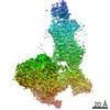 12746MC M: map data used to model this data C: citing same article ( |
|---|---|
| Similar structure data |


-Links
 PDBj
PDBj


- Assembly
| Deposited unit | 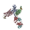
|
|---|---|
| 1 |
|
-Components
-Guanine nucleotide-binding protein ... , 3 types, 3 molecules ABG
| #1: Protein | Mass: 40415.031 Da / Num. of mol.: 1 Source method: isolated from a genetically manipulated source Source: (gene. exp.) Homo sapiens (human) / Gene: GNAI1 / Production host:  Escherichia coli BL21(DE3) (bacteria) / References: UniProt: P63096 Escherichia coli BL21(DE3) (bacteria) / References: UniProt: P63096 |
|---|---|
| #2: Protein | Mass: 37416.930 Da / Num. of mol.: 1 / Source method: isolated from a natural source / Source: (natural) Bos taurus (cattle) / References: UniProt: P62871 |
| #4: Protein | Mass: 8556.918 Da / Num. of mol.: 1 / Source method: isolated from a natural source / Details: UNP P02698 / Source: (natural) Bos taurus (cattle) / References: UniProt: P02698 |
-Protein , 2 types, 2 molecules CI
| #3: Protein | Mass: 42726.168 Da / Num. of mol.: 1 Source method: isolated from a genetically manipulated source Details: UNP P51681 / Source: (gene. exp.) Homo sapiens (human) / Gene: CCR5, CMKBR5 / Production host:   Spodoptera frugiperda (fall armyworm) / References: UniProt: P51681 Spodoptera frugiperda (fall armyworm) / References: UniProt: P51681 |
|---|---|
| #5: Protein | Chemokine / EoCP / Eosinophil chemotactic cytokine / SIS-delta / Small-inducible cytokine A5 / T cell-specific ...EoCP / Eosinophil chemotactic cytokine / SIS-delta / Small-inducible cytokine A5 / T cell-specific protein P228 / TCP228 / T-cell-specific protein RANTES Mass: 7857.123 Da / Num. of mol.: 1 Source method: isolated from a genetically manipulated source Details: UNP P13501 / Source: (gene. exp.) Homo sapiens (human) / Gene: CCL5, D17S136E, SCYA5 / Production host: Escherichia coli (E. coli) / References: UniProt: P13501 |
-Antibody , 2 types, 2 molecules HF
| #6: Antibody | Mass: 23542.248 Da / Num. of mol.: 1 Source method: isolated from a genetically manipulated source Source: (gene. exp.) Mus musculus (house mouse) / Production host: Mus musculus (house mouse) |
|---|---|
| #7: Antibody | Mass: 23921.592 Da / Num. of mol.: 1 Source method: isolated from a genetically manipulated source Source: (gene. exp.) Mus musculus (house mouse) / Production host: Mus musculus (house mouse) |
-Details
| Has ligand of interest | N |
|---|
-Experimental details
-Experiment
| Experiment | Method: ELECTRON MICROSCOPY |
|---|---|
| EM experiment | Aggregation state: PARTICLE / 3D reconstruction method: single particle reconstruction |
- Sample preparation
| Component |
| ||||||||||||||||||||||||||||||||||||||||||||||||
|---|---|---|---|---|---|---|---|---|---|---|---|---|---|---|---|---|---|---|---|---|---|---|---|---|---|---|---|---|---|---|---|---|---|---|---|---|---|---|---|---|---|---|---|---|---|---|---|---|---|
| Molecular weight | Units: KILODALTONS/NANOMETER / Experimental value: NO | ||||||||||||||||||||||||||||||||||||||||||||||||
| Source (natural) |
| ||||||||||||||||||||||||||||||||||||||||||||||||
| Source (recombinant) |
| ||||||||||||||||||||||||||||||||||||||||||||||||
| Buffer solution | pH: 7.5 | ||||||||||||||||||||||||||||||||||||||||||||||||
| Buffer component |
| ||||||||||||||||||||||||||||||||||||||||||||||||
| Specimen | Conc.: 2.5 mg/ml / Embedding applied: NO / Shadowing applied: NO / Staining applied: NO / Vitrification applied: YES | ||||||||||||||||||||||||||||||||||||||||||||||||
| Specimen support | Grid material: COPPER / Grid mesh size: 200 divisions/in. / Grid type: Quantifoil R1.2/1.3 | ||||||||||||||||||||||||||||||||||||||||||||||||
| Vitrification | Instrument: FEI VITROBOT MARK IV / Cryogen name: ETHANE / Humidity: 100 % / Chamber temperature: 277 K |
- Electron microscopy imaging
| Microscopy | Model: TFS GLACIOS |
|---|---|
| Electron gun | Electron source: FIELD EMISSION GUN / Accelerating voltage: 200 kV / Illumination mode: FLOOD BEAM |
| Electron lens | Mode: BRIGHT FIELDBright-field microscopy |
| Image recording | Electron dose: 49 e/Å2 / Film or detector model: GATAN K3 (6k x 4k) / Num. of grids imaged: 1 / Num. of real images: 2466 |
- Processing
| Software | Name: PHENIX / Version: 1.19_4092: / Classification: refinement | ||||||||||||||||||||||||||||||||||||||||
|---|---|---|---|---|---|---|---|---|---|---|---|---|---|---|---|---|---|---|---|---|---|---|---|---|---|---|---|---|---|---|---|---|---|---|---|---|---|---|---|---|---|
| EM software |
| ||||||||||||||||||||||||||||||||||||||||
| CTF correction | Type: NONE | ||||||||||||||||||||||||||||||||||||||||
| 3D reconstruction | Resolution: 3.15 Å / Resolution method: FSC 0.143 CUT-OFF / Num. of particles: 345458 / Algorithm: FOURIER SPACE / Symmetry type: POINT | ||||||||||||||||||||||||||||||||||||||||
| Atomic model building |
| ||||||||||||||||||||||||||||||||||||||||
| Atomic model building |
| ||||||||||||||||||||||||||||||||||||||||
| Refine LS restraints |
|