- PDB-7o45: Crystal structure of ADD domain of the human DNMT3B methyltransferase -
+
Open data
ID or keywords:
Loading...
-
Basic information
Entry
Database: PDB / ID: 7o45
Title
Crystal structure of ADD domain of the human DNMT3B methyltransferase
Components
Isoform 6 of DNA (cytosine-5)-methyltransferase 3B
Keywords
TRANSFERASE / Methyltransferase 3B / DNA methylation / DNMT3B
Function / homology
Function and homology information
: / DNA (cytosine-5-)-methyltransferase activity, acting on CpG substrates / DNA-methyltransferase activity / DNA (cytosine-5-)-methyltransferase / DNA (cytosine-5-)-methyltransferase activity / SUMOylation of DNA methylation proteins / : / catalytic complex / DNA methylation / PRC2 methylates histones and DNA ...: / DNA (cytosine-5-)-methyltransferase activity, acting on CpG substrates / DNA-methyltransferase activity / DNA (cytosine-5-)-methyltransferase / DNA (cytosine-5-)-methyltransferase activity / SUMOylation of DNA methylation proteins / : / catalytic complex / DNA methylation / PRC2 methylates histones and DNA / Defective pyroptosis / NoRC negatively regulates rRNA expression / transcription corepressor activity / positive regulation of gene expression / negative regulation of transcription by RNA polymerase II / DNA binding / nucleoplasm / metal ion binding / nucleus Similarity search - Function
A: Isoform 6 of DNA (cytosine-5)-methyltransferase 3B B: Isoform 6 of DNA (cytosine-5)-methyltransferase 3B C: Isoform 6 of DNA (cytosine-5)-methyltransferase 3B D: Isoform 6 of DNA (cytosine-5)-methyltransferase 3B hetero molecules
A: Isoform 6 of DNA (cytosine-5)-methyltransferase 3B B: Isoform 6 of DNA (cytosine-5)-methyltransferase 3B C: Isoform 6 of DNA (cytosine-5)-methyltransferase 3B hetero molecules
Component-ID: 0 / Beg auth comp-ID: ASP / Beg label comp-ID: ASP / End auth comp-ID: THR / End label comp-ID: THR / Refine code: 0 / Auth seq-ID: 414 - 554 / Label seq-ID: 6 - 146
Dom-ID
Ens-ID
Auth asym-ID
Label asym-ID
1
1
A
A
2
1
B
B
1
2
A
A
2
2
C
C
1
3
B
B
2
3
C
C
NCS ensembles :
ID
1
2
3
-
Components
#1: Protein
Isoform6ofDNA (cytosine-5)-methyltransferase3B / Dnmt3b / DNA methyltransferase HsaIIIB / DNA MTase HsaIIIB / M.HsaIIIB
Mass: 16556.688 Da / Num. of mol.: 4 Source method: isolated from a genetically manipulated source Source: (gene. exp.) Homo sapiens (human) / Gene: DNMT3B / Production host: Escherichia coli (E. coli) References: UniProt: Q9UBC3, DNA (cytosine-5-)-methyltransferase
Resolution: 2.1→64.38 Å / Cor.coef. Fo:Fc: 0.956 / Cor.coef. Fo:Fc free: 0.943 / SU B: 9.535 / SU ML: 0.131 / SU R Cruickshank DPI: 0.2062 / Cross valid method: THROUGHOUT / σ(F): 0 / ESU R: 0.206 / ESU R Free: 0.171 / Stereochemistry target values: MAXIMUM LIKELIHOOD Details: HYDROGENS HAVE BEEN ADDED IN THE RIDING POSITIONS U VALUES : WITH TLS ADDED
Rfactor
Num. reflection
% reflection
Selection details
Rfree
0.2281
1810
5 %
RANDOM
Rwork
0.2
-
-
-
obs
0.2015
34698
92.05 %
-
Solvent computation
Ion probe radii: 0.7 Å / Shrinkage radii: 0.7 Å / VDW probe radii: 1 Å / Solvent model: MASK
In the structure databanks used in Yorodumi, some data are registered as the other names, "COVID-19 virus" and "2019-nCoV". Here are the details of the virus and the list of structure data.
Jan 31, 2019. EMDB accession codes are about to change! (news from PDBe EMDB page)
EMDB accession codes are about to change! (news from PDBe EMDB page)
The allocation of 4 digits for EMDB accession codes will soon come to an end. Whilst these codes will remain in use, new EMDB accession codes will include an additional digit and will expand incrementally as the available range of codes is exhausted. The current 4-digit format prefixed with “EMD-” (i.e. EMD-XXXX) will advance to a 5-digit format (i.e. EMD-XXXXX), and so on. It is currently estimated that the 4-digit codes will be depleted around Spring 2019, at which point the 5-digit format will come into force.
The EM Navigator/Yorodumi systems omit the EMD- prefix.
Related info.:Q: What is EMD? / ID/Accession-code notation in Yorodumi/EM Navigator
Yorodumi is a browser for structure data from EMDB, PDB, SASBDB, etc.
This page is also the successor to EM Navigator detail page, and also detail information page/front-end page for Omokage search.
The word "yorodu" (or yorozu) is an old Japanese word meaning "ten thousand". "mi" (miru) is to see.
Related info.:EMDB / PDB / SASBDB / Comparison of 3 databanks / Yorodumi Search / Aug 31, 2016. New EM Navigator & Yorodumi / Yorodumi Papers / Jmol/JSmol / Function and homology information / Changes in new EM Navigator and Yorodumi
 Movie
Movie Controller

 Yorodumi
 Open data
 Basic information
 Components
 Keywords
 TRANSFERASE / Methyltransferase 3B /
  Function and homology information
 Authors
 Russian Federation, 1items
  Citation
 Structure visualization
 Downloads & links
 Other downloads
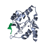
 PDBj
PDBj


 Assembly


 Mass: 65.409 Da / Num. of mol.: 12 / Source method: obtained synthetically / Formula: Zn / Feature type: SUBJECT OF INVESTIGATION
 Mass: 79.904 Da / Num. of mol.: 3 / Source method: obtained synthetically / Formula: Br
 Mass: 18.015 Da / Num. of mol.: 129 / Source method: isolated from a natural source / Formula: H2O
 Sample preparation
 / Beamline: MASSIF-3 / Wavelength: 1.284 Å
 Processing