 Movie
Movie Controller

[English] 日本語
 Yorodumi
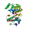
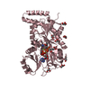
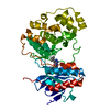
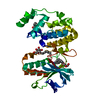
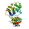
- PDB-7o0e: Crystal structure of GH30 (mutant E188A) complexed with aldotriur... -
+ Open data

- Basic information
| Entry | Database: PDB / ID: 7o0e | ||||||
|---|---|---|---|---|---|---|---|
| Title | Crystal structure of GH30 (mutant E188A) complexed with aldotriuronic acid from Thermothelomyces thermophila. | ||||||
 Components Components | GH30 family xylanase | ||||||
 Keywords Keywords |  HYDROLASE / xylanase / GH30 / subfamily 7 / fungal HYDROLASE / xylanase / GH30 / subfamily 7 / fungal | ||||||
| Function / homology |  Function and homology informationglucosylceramidase activity / sphingolipid metabolic process / Hydrolases; Glycosylases; Glycosidases, i.e. enzymes that hydrolyse O- and S-glycosyl compounds / extracellular region Function and homology informationglucosylceramidase activity / sphingolipid metabolic process / Hydrolases; Glycosylases; Glycosidases, i.e. enzymes that hydrolyse O- and S-glycosyl compounds / extracellular regionSimilarity search - Function | ||||||
| Biological species |  Myceliophthora thermophila (fungus) Myceliophthora thermophila (fungus) | ||||||
| Method | X-RAY DIFFRACTION / SYNCHROTRON / MOLECULAR REPLACEMENT / Resolution: 1.85 Å | ||||||
 Authors Authors | Dimarogona, M. / Kosinas, C. / Feiler, C. / Weiss, M.S. / Topakas, E. / Nikolaivits, E. | ||||||
 Citation Citation | Journal: Carbohydrate Polymers / Year: 2021 Title: Unique features of the bifunctional GH30 from Thermothelomyces thermophila revealed by structural and mutational studies Authors: Nikolaivits, E. / Pentari, C. / Kosinas, C. / Feiler, C.G. / Spiliopoulou, M. / Weiss, M.S. / Dimarogona, M. / Topakas, E. | ||||||
| History |
|
- Structure visualization
| Structure viewer | Molecule: MolmilJmol/JSmol |
|---|
- Downloads & links
-Download
| PDBx/mmCIF format | 7o0e.cif.gz | 357 KB | Display | PDBx/mmCIF format |
|---|---|---|---|---|
| PDB format | pdb7o0e.ent.gz | 288.3 KB | Display | PDB format |
| PDBx/mmJSON format | 7o0e.json.gz | Tree view | PDBx/mmJSON format | |
| Others |  Other downloads Other downloads |
-Validation report
| Arichive directory | https://data.pdbj.org/pub/pdb/validation_reports/o0/7o0eftp://data.pdbj.org/pub/pdb/validation_reports/o0/7o0e | HTTPS FTP |
|---|
-Related structure data
| Related structure data | 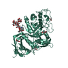 7ncxSC S: Starting model for refinement C: citing same article ( |
|---|---|
| Similar structure data |


-Links
 PDBj
PDBj

- Assembly
| Deposited unit | 
| ||||||||
|---|---|---|---|---|---|---|---|---|---|
| 1 | 
| ||||||||
| 2 | 
| ||||||||
| Unit cell |
|
-Components
-Protein , 1 types, 2 molecules GA
| #1: Protein | Mass: 48228.109 Da / Num. of mol.: 2 / Mutation: E188A Source method: isolated from a genetically manipulated source Source: (gene. exp.) Myceliophthora thermophila (strain ATCC 42464 / BCRC 31852 / DSM 1799) (fungus)Strain: ATCC 42464 / BCRC 31852 / DSM 1799 / Gene: Xyn30A, MYCTH_38558 / Production host: Komagataella pastoris (fungus)References: UniProt: G2Q1N4, Hydrolases; Glycosylases; Glycosidases, i.e. enzymes that hydrolyse O- and S-glycosyl compounds |
|---|
-Sugars , 2 types, 3 molecules
| #2: Polysaccharide | alpha-D-mannopyranose-(1-3)-beta-D-mannopyranose-(1-4)-2-acetamido-2-deoxy-beta-D-glucopyranose-(1- ...alpha-D-mannopyranose-(1-3)-beta-D-mannopyranose-(1-4)-2-acetamido-2-deoxy-beta-D-glucopyranose-(1-4)-2-acetamido-2-deoxy-beta-D-glucopyranose / Mass: 748.682 Da / Num. of mol.: 1 Source method: isolated from a genetically manipulated source |
|---|---|
| #3: Polysaccharide | Type: oligosaccharide / Mass: 472.396 Da / Num. of mol.: 2 / Source method: obtained synthetically |
-Non-polymers , 4 types, 693 molecules 


| #4: Chemical | ChemComp-GLY / Glycine Type: peptide linking / Mass: 75.067 Da / Num. of mol.: 5 / Source method: obtained synthetically / Formula: C2H5NO2 Type: peptide linking / Mass: 75.067 Da / Num. of mol.: 5 / Source method: obtained synthetically / Formula: C2H5NO2#5: Chemical | ChemComp-EDO / Ethylene glycol Mass: 62.068 Da / Num. of mol.: 4 / Source method: obtained synthetically / Formula: C2H6O2 Mass: 62.068 Da / Num. of mol.: 4 / Source method: obtained synthetically / Formula: C2H6O2#6: Chemical | ChemComp-PEG / | Diethylene glycol Mass: 106.120 Da / Num. of mol.: 1 / Source method: obtained synthetically / Formula: C4H10O3 Mass: 106.120 Da / Num. of mol.: 1 / Source method: obtained synthetically / Formula: C4H10O3#7: Water | ChemComp-HOH / | WaterMass: 18.015 Da / Num. of mol.: 683 / Source method: isolated from a natural source / Formula: H2O |
|---|
-Details
| Has ligand of interest | Y |
|---|
-Experimental details
-Experiment
| Experiment | Method: X-RAY DIFFRACTION / Number of used crystals: 1 |
|---|
- Sample preparation
| Crystal | Density Matthews: 2.02013588 Å3/Da / Density % sol: 39.1508331 % |
|---|---|
| Crystal grow | Temperature: 293 K / Method: vapor diffusion, sitting drop / Details: 0.1 M SPG pH 7.0, 25% w/v PEG 1500 |
-Data collection
| Diffraction | Mean temperature: 100 K / Serial crystal experiment: N |
|---|---|
| Diffraction source | Source: SYNCHROTRON / Site: BESSY  / Beamline: 14.2 / Wavelength: 0.9184 Å / Beamline: 14.2 / Wavelength: 0.9184 Å |
| Detector | Type: DECTRIS PILATUS3 S 2M / Detector: PIXEL / Date: Feb 3, 2021 |
| Radiation | Protocol: SINGLE WAVELENGTH / Monochromatic (M) / Laue (L): M / Scattering type: x-ray |
| Radiation wavelength | Wavelength: 0.9184 Å / Relative weight: 1 |
| Reflection | Resolution: 1.85→46.884 Å / Num. obs: 67654 / % possible obs: 100 % / Redundancy: 6.7 % / CC1/2: 0.997 / Net I/σ(I): 10.5 |
| Reflection shell | Resolution: 1.85→1.89 Å / Num. unique obs: 4130 / CC1/2: 0.564 |
- Processing
| Software |
| |||||||||||||||||||||||||||||||||||||||||||||||||||||||||||||||||||||||||||
|---|---|---|---|---|---|---|---|---|---|---|---|---|---|---|---|---|---|---|---|---|---|---|---|---|---|---|---|---|---|---|---|---|---|---|---|---|---|---|---|---|---|---|---|---|---|---|---|---|---|---|---|---|---|---|---|---|---|---|---|---|---|---|---|---|---|---|---|---|---|---|---|---|---|---|---|---|
| Refinement | Method to determine structure: MOLECULAR REPLACEMENT Starting model: 7NCX Resolution: 1.85→46.88 Å / Cor.coef. Fo:Fc: 0.968 / Cor.coef. Fo:Fc free: 0.955 / SU B: 6.581 / SU ML: 0.097 / Cross valid method: THROUGHOUT / σ(F): 0 / ESU R: 0.139 / ESU R Free: 0.124 / Stereochemistry target values: MAXIMUM LIKELIHOOD Details: HYDROGENS HAVE BEEN ADDED IN THE RIDING POSITIONS U VALUES : WITH TLS ADDED
| |||||||||||||||||||||||||||||||||||||||||||||||||||||||||||||||||||||||||||
| Solvent computation | Ion probe radii: 0.8 Å / Shrinkage radii: 0.8 Å / VDW probe radii: 1.2 Å / Solvent model: MASK | |||||||||||||||||||||||||||||||||||||||||||||||||||||||||||||||||||||||||||
| Displacement parameters | Biso max: 79.22 Å2 / Biso mean: 21.117 Å2 / Biso min: 11.59 Å2
| |||||||||||||||||||||||||||||||||||||||||||||||||||||||||||||||||||||||||||
| Refinement step | Cycle: final / Resolution: 1.85→46.88 Å
| |||||||||||||||||||||||||||||||||||||||||||||||||||||||||||||||||||||||||||
| Refine LS restraints |
| |||||||||||||||||||||||||||||||||||||||||||||||||||||||||||||||||||||||||||
| LS refinement shell | Resolution: 1.85→1.898 Å / Rfactor Rfree error: 0 / Total num. of bins used: 20
| |||||||||||||||||||||||||||||||||||||||||||||||||||||||||||||||||||||||||||
| Refinement TLS params. | Method: refined / Refine-ID: X-RAY DIFFRACTION
| |||||||||||||||||||||||||||||||||||||||||||||||||||||||||||||||||||||||||||
| Refinement TLS group |
|
