Movie
Movie Controller


+ Open data

- Basic information
| Entry | Database: PDB / ID: 7nyc | ||||||||||||
|---|---|---|---|---|---|---|---|---|---|---|---|---|---|
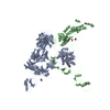
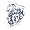
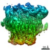
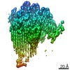
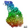
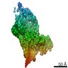
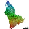
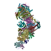
| Title | cryoEM structure of 3C9-sMAC | ||||||||||||
 Components Components |
| ||||||||||||
 Keywords Keywords |  IMMUNE SYSTEM / Complement / MACPF / Membrane Attack Complex / CDC / pore forming IMMUNE SYSTEM / Complement / MACPF / Membrane Attack Complex / CDC / pore forming | ||||||||||||
| Function / homology |  Function and homology information Function and homology informationcell killing / Terminal pathway of complement / membrane attack complex / complement binding / other organism cell membrane / Activation of C3 and C5 / negative regulation of macrophage chemotaxis / complement activation, alternative pathway / complement activation / chemokine activity ...cell killing / Terminal pathway of complement / membrane attack complex / complement binding / other organism cell membrane / Activation of C3 and C5 / negative regulation of macrophage chemotaxis / complement activation, alternative pathway / complement activation / chemokine activity / retinol binding / endopeptidase inhibitor activity / positive regulation of vascular endothelial growth factor production / complement activation, classical pathway / positive regulation of chemokine production / Peptide ligand-binding receptors / Regulation of Complement cascade / protein homooligomerization / extracellular vesicle / chemotaxis / positive regulation of immune response / G alpha (i) signalling events / blood microparticle / killing of cells of another organism / in utero embryonic development / cell surface receptor signaling pathway / immune response / inflammatory response / G protein-coupled receptor signaling pathway / signaling receptor binding / innate immune response / protein-containing complex binding / extracellular space / extracellular exosome / extracellular region / membrane / plasma membraneSimilarity search - Function | ||||||||||||
| Biological species |  Homo sapiens (human) Homo sapiens (human) | ||||||||||||
| Method | ELECTRON MICROSCOPY / single particle reconstruction / cryo EM / Resolution: 3.54 Å | ||||||||||||
 Authors Authors | Menny, A. / Couves, E.C. / Bubeck, D. | ||||||||||||
| Funding support | European Union,  United Kingdom, 3items United Kingdom, 3items
| ||||||||||||
 Citation Citation | Journal: Nat Commun / Year: 2021 Title: Structural basis of soluble membrane attack complex packaging for clearance. Authors: Anaïs Menny / Marie V Lukassen / Emma C Couves / Vojtech Franc / Albert J R Heck / Doryen Bubeck /  Abstract: Unregulated complement activation causes inflammatory and immunological pathologies with consequences for human disease. To prevent bystander damage during an immune response, extracellular ...Unregulated complement activation causes inflammatory and immunological pathologies with consequences for human disease. To prevent bystander damage during an immune response, extracellular chaperones (clusterin and vitronectin) capture and clear soluble precursors to the membrane attack complex (sMAC). However, how these chaperones block further polymerization of MAC and prevent the complex from binding target membranes remains unclear. Here, we address that question by combining cryo electron microscopy (cryoEM) and cross-linking mass spectrometry (XL-MS) to solve the structure of sMAC. Together our data reveal how clusterin recognizes and inhibits polymerizing complement proteins by binding a negatively charged surface of sMAC. Furthermore, we show that the pore-forming C9 protein is trapped in an intermediate conformation whereby only one of its two transmembrane β-hairpins has unfurled. This structure provides molecular details for immune pore formation and helps explain a complement control mechanism that has potential implications for how cell clearance pathways mediate immune homeostasis. | ||||||||||||
| History |
|
- Structure visualization
| Movie |
Movie viewer |
|---|---|
| Structure viewer | Molecule: MolmilJmol/JSmol |
- Downloads & links
-Download
| PDBx/mmCIF format | 7nyc.cif.gz | 1 MB | Display | PDBx/mmCIF format |
|---|---|---|---|---|
| PDB format | pdb7nyc.ent.gz | 843.7 KB | Display | PDB format |
| PDBx/mmJSON format | 7nyc.json.gz | Tree view | PDBx/mmJSON format | |
| Others |  Other downloads Other downloads |
-Validation report
| Arichive directory | https://data.pdbj.org/pub/pdb/validation_reports/ny/7nycftp://data.pdbj.org/pub/pdb/validation_reports/ny/7nyc | HTTPS FTP |
|---|
-Related structure data
| Related structure data | 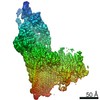 12650MC 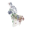 7nydC C: citing same article ( M: map data used to model this data |
|---|---|
| Similar structure data |


-Links
 PDBj
PDBj


- Assembly
| Deposited unit | 
|
|---|---|
| 1 |
|
-Components
-Complement component ... , 6 types, 8 molecules CDEGHIBF
| #1: Protein | Mass: 91221.484 Da / Num. of mol.: 1 / Source method: isolated from a natural source / Source: (natural) Homo sapiens (human) / References: UniProt: P10643 | ||||
|---|---|---|---|---|---|
| #2: Protein | Mass: 61122.852 Da / Num. of mol.: 1 / Source method: isolated from a natural source / Source: (natural) Homo sapiens (human) / References: UniProt: P07358 | ||||
| #3: Protein | Mass: 61782.992 Da / Num. of mol.: 1 / Source method: isolated from a natural source / Source: (natural) Homo sapiens (human) / References: UniProt: P07357 | ||||
| #4: Protein | Mass: 61056.594 Da / Num. of mol.: 3 / Source method: isolated from a natural source / Source: (natural) Homo sapiens (human) / References: UniProt: P02748#5: Protein | | Mass: 102541.312 Da / Num. of mol.: 1 / Source method: isolated from a natural source / Source: (natural) Homo sapiens (human) / References: UniProt: P13671#7: Protein | | Mass: 20410.105 Da / Num. of mol.: 1 / Source method: isolated from a natural source / Source: (natural) Homo sapiens (human) / References: UniProt: P07360 |
-Protein / Non-polymers , 2 types, 4 molecules A
| #11: Chemical |  Mass: 40.078 Da / Num. of mol.: 3 / Source method: obtained synthetically / Formula: Ca Mass: 40.078 Da / Num. of mol.: 3 / Source method: obtained synthetically / Formula: Ca#6: Protein | | Complement component 5 / C3 and PZP-like alpha-2-macroglobulin domain-containing protein 4Mass: 186546.656 Da / Num. of mol.: 1 / Source method: isolated from a natural source / Source: (natural) Homo sapiens (human) / References: UniProt: P01031 |
|---|
-Sugars , 5 types, 14 molecules 


| #8: Polysaccharide | 2-acetamido-2-deoxy-beta-D-glucopyranose-(1-4)-2-acetamido-2-deoxy-beta-D-glucopyranose / Mass: 424.401 Da / Num. of mol.: 1 Source method: isolated from a genetically manipulated source | ||||
|---|---|---|---|---|---|
| #9: Polysaccharide | beta-D-mannopyranose-(1-4)-2-acetamido-2-deoxy-beta-D-glucopyranose-(1-4)-2-acetamido-2-deoxy-beta- ...beta-D-mannopyranose-(1-4)-2-acetamido-2-deoxy-beta-D-glucopyranose-(1-4)-2-acetamido-2-deoxy-beta-D-glucopyranose / Mass: 586.542 Da / Num. of mol.: 1 Source method: isolated from a genetically manipulated source | ||||
| #10: Sugar | ChemComp-BMA / Mannose Type: D-saccharide, beta linking / Mass: 180.156 Da / Num. of mol.: 7 / Source method: obtained synthetically / Formula: C6H12O6 Type: D-saccharide, beta linking / Mass: 180.156 Da / Num. of mol.: 7 / Source method: obtained synthetically / Formula: C6H12O6#12: Sugar | ChemComp-NAG / N-Acetylglucosamine Type: D-saccharide, beta linking / Mass: 221.208 Da / Num. of mol.: 4 / Source method: obtained synthetically / Formula: C8H15NO6 Type: D-saccharide, beta linking / Mass: 221.208 Da / Num. of mol.: 4 / Source method: obtained synthetically / Formula: C8H15NO6#13: Sugar | ChemComp-FUC / | Fucose Type: L-saccharide, alpha linking / Mass: 164.156 Da / Num. of mol.: 1 / Source method: obtained synthetically / Formula: C6H12O5 Type: L-saccharide, alpha linking / Mass: 164.156 Da / Num. of mol.: 1 / Source method: obtained synthetically / Formula: C6H12O5 |
-Details
| Has ligand of interest | N |
|---|
-Experimental details
-Experiment
| Experiment | Method: ELECTRON MICROSCOPY |
|---|---|
| EM experiment | Aggregation state: PARTICLE / 3D reconstruction method: single particle reconstruction |
- Sample preparation
| Component | Name: 3C9-sMAC / Type: COMPLEX Details: This sMAC oligomer contains one copy of C5b8 and 3 copies of C9, as well as multiple copies of the chaperones vitronectin and clusterin Entity ID: #1-#7 / Source: NATURAL |
|---|---|
| Molecular weight | Value: 1 MDa / Experimental value: YES |
| Source (natural) | Organism: Homo sapiens (human) |
| Buffer solution | pH: 7.4 |
| Specimen | Conc.: 0.065 mg/ml / Embedding applied: NO / Shadowing applied: NO / Staining applied: NO / Vitrification applied: YES |
| Vitrification | Instrument: FEI VITROBOT MARK III / Cryogen name: ETHANE / Humidity: 95 % / Chamber temperature: 295 K |
- Electron microscopy imaging
| Experimental equipment |  Model: Titan Krios / Image courtesy: FEI Company |
|---|---|
| Microscopy | Model: FEI TITAN KRIOS |
| Electron gun | Electron source: FIELD EMISSION GUN / Accelerating voltage: 300 kV / Illumination mode: FLOOD BEAM |
| Electron lens | Mode: BRIGHT FIELDBright-field microscopy / C2 aperture diameter: 70 µm |
| Image recording | Electron dose: 40 e/Å2 / Detector mode: COUNTING / Film or detector model: GATAN K2 QUANTUM (4k x 4k) |
- Processing
| Software | Name: REFMAC / Version: 5.8.0253 / Classification: refinement | ||||||||||||||||||||||||||||||||||||||||||||||||||||||||||||||||||||||||||||||||||||||||||||||||||||||||||
|---|---|---|---|---|---|---|---|---|---|---|---|---|---|---|---|---|---|---|---|---|---|---|---|---|---|---|---|---|---|---|---|---|---|---|---|---|---|---|---|---|---|---|---|---|---|---|---|---|---|---|---|---|---|---|---|---|---|---|---|---|---|---|---|---|---|---|---|---|---|---|---|---|---|---|---|---|---|---|---|---|---|---|---|---|---|---|---|---|---|---|---|---|---|---|---|---|---|---|---|---|---|---|---|---|---|---|---|
| EM software |
| ||||||||||||||||||||||||||||||||||||||||||||||||||||||||||||||||||||||||||||||||||||||||||||||||||||||||||
| CTF correction | Type: PHASE FLIPPING AND AMPLITUDE CORRECTION | ||||||||||||||||||||||||||||||||||||||||||||||||||||||||||||||||||||||||||||||||||||||||||||||||||||||||||
| 3D reconstruction | Resolution: 3.54 Å / Resolution method: FSC 0.143 CUT-OFF / Num. of particles: 85151 / Symmetry type: POINT | ||||||||||||||||||||||||||||||||||||||||||||||||||||||||||||||||||||||||||||||||||||||||||||||||||||||||||
| Atomic model building | Protocol: RIGID BODY FIT | ||||||||||||||||||||||||||||||||||||||||||||||||||||||||||||||||||||||||||||||||||||||||||||||||||||||||||
| Atomic model building |
| ||||||||||||||||||||||||||||||||||||||||||||||||||||||||||||||||||||||||||||||||||||||||||||||||||||||||||
| Refinement | Resolution: 3.5→418.8 Å / Cor.coef. Fo:Fc: 0.891 / SU B: 16.753 / SU ML: 0.236 / ESU R: 0.166 Stereochemistry target values: MAXIMUM LIKELIHOOD WITH PHASES
| ||||||||||||||||||||||||||||||||||||||||||||||||||||||||||||||||||||||||||||||||||||||||||||||||||||||||||
| Solvent computation | Solvent model: PARAMETERS FOR MASK CACLULATION | ||||||||||||||||||||||||||||||||||||||||||||||||||||||||||||||||||||||||||||||||||||||||||||||||||||||||||
| Displacement parameters | Biso mean: 268.658 Å2
| ||||||||||||||||||||||||||||||||||||||||||||||||||||||||||||||||||||||||||||||||||||||||||||||||||||||||||
| Refinement step | Cycle: 1 / Total: 41391 | ||||||||||||||||||||||||||||||||||||||||||||||||||||||||||||||||||||||||||||||||||||||||||||||||||||||||||
| Refine LS restraints |
|