 Movie
Movie Controller

[English] 日本語
 Yorodumi
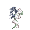
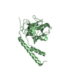
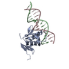
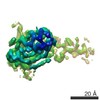
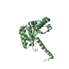
- PDB-2wcy: NMR solution structure of factor I-like modules of complement C7. -
+ Open data

- Basic information
| Entry | Database: PDB / ID: 2wcy | ||||||
|---|---|---|---|---|---|---|---|
| Title | NMR solution structure of factor I-like modules of complement C7. | ||||||
 Components Components | COMPLEMENT COMPONENT C7 | ||||||
 Keywords Keywords |  IMMUNE SYSTEM / DISULFIDE BOND / IMMUNE RESPONSE / FACTOR I MODULE / C7 / FIM / EGF / MAC / FOLN / SUSHI / FIMAC / KAZAL / COMPLEMENT ALTERNATE PATHWAY / FOLLISTATIN / POLYMORPHISM / GLYCOPROTEIN / SECRETED / DISULFIDE / CYTOLYSIS / COMPLEMENT / COMPLEMENT PATHWAY / MEMBRANE ATTACK COMPLEX / INNATE IMMUNITY / EGF-LIKE DOMAIN / DISEASE MUTATION IMMUNE SYSTEM / DISULFIDE BOND / IMMUNE RESPONSE / FACTOR I MODULE / C7 / FIM / EGF / MAC / FOLN / SUSHI / FIMAC / KAZAL / COMPLEMENT ALTERNATE PATHWAY / FOLLISTATIN / POLYMORPHISM / GLYCOPROTEIN / SECRETED / DISULFIDE / CYTOLYSIS / COMPLEMENT / COMPLEMENT PATHWAY / MEMBRANE ATTACK COMPLEX / INNATE IMMUNITY / EGF-LIKE DOMAIN / DISEASE MUTATION | ||||||
| Function / homology |  Function and homology information Function and homology informationTerminal pathway of complement / membrane attack complex / complement activation, alternative pathway / complement activation / complement activation, classical pathway / Regulation of Complement cascade / positive regulation of immune response / killing of cells of another organism / extracellular space / extracellular exosome ...Terminal pathway of complement / membrane attack complex / complement activation, alternative pathway / complement activation / complement activation, classical pathway / Regulation of Complement cascade / positive regulation of immune response / killing of cells of another organism / extracellular space / extracellular exosome / extracellular region / plasma membraneSimilarity search - Function | ||||||
| Biological species |  HOMO SAPIENS (human) HOMO SAPIENS (human) | ||||||
| Method | SOLUTION NMR / SIMULATED ANNEAILING, RESTRAINED MOLECULAR DYNAMICS | ||||||
 Authors Authors | Phelan, M.M. / Thai, C.T. / Soares, D.C. / Ogata, R.T. / Barlow, P.N. / Bramham, J. | ||||||
 Citation Citation | Journal: J.Biol.Chem. / Year: 2009 Title: Solution Structure of Factor I-Like Modules from Complement C7 Reveals a Pair of Follistatin Domains in Compact Pseudosymmetric Arrangement. Authors: Phelan, M.M. / Thai, C.T. / Soares, D.C. / Ogata, R.T. / Barlow, P.N. / Bramham, J. #1: Journal: Biomol. NMR Assign. / Year: 2009 Title: 1H, 15N and 13C Resonance Assignment of the Pair of Factor-I Like Modules of the Complement Protein C7 Authors: Phelan, M.M. / Thai, C.T. / Herbert, A.P. / Bella, J. / Uhrin, D. / Ogata, R.T. / Barlow, P.N. / Bramham, J. | ||||||
| History |
| ||||||
| Remark 650 | HELIX DETERMINATION METHOD: AUTHOR PROVIDED. | ||||||
| Remark 700 | SHEET DETERMINATION METHOD: AUTHOR PROVIDED. |
- Structure visualization
| Structure viewer | Molecule: MolmilJmol/JSmol |
|---|
- Downloads & links
-Download
| PDBx/mmCIF format | 2wcy.cif.gz | 2.1 MB | Display | PDBx/mmCIF format |
|---|---|---|---|---|
| PDB format | pdb2wcy.ent.gz | 1.8 MB | Display | PDB format |
| PDBx/mmJSON format | 2wcy.json.gz | Tree view | PDBx/mmJSON format | |
| Others |  Other downloads Other downloads |
-Validation report
| Arichive directory | https://data.pdbj.org/pub/pdb/validation_reports/wc/2wcyftp://data.pdbj.org/pub/pdb/validation_reports/wc/2wcy | HTTPS FTP |
|---|
-Related structure data
| Similar structure data | |
|---|---|
| Other databases |


-Links
 PDBj
PDBj


- Assembly
| Deposited unit | 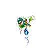
| |||||||||
|---|---|---|---|---|---|---|---|---|---|---|
| 1 |
| |||||||||
| NMR ensembles |
|
-Components
| #1: Protein | Mass: 16977.494 Da / Num. of mol.: 1 / Fragment: FACTOR I-LIKE MODULES (FIMS), RESIDUES 693-843 Source method: isolated from a genetically manipulated source Details: N TERMINAL CLONING ARTEFACT GSHM / Source: (gene. exp.) HOMO SAPIENS (human) / Tissue: BLOOD / Description: COMPONENT OF HUMAN COMPLEMENT SYSTEM / Cellular location: EXTRACELLULARGlossary of biology / Production host:  ESCHERICHIA COLI (E. coli) / Strain (production host): ORIGAMI B / References: UniProt: P10643 ESCHERICHIA COLI (E. coli) / Strain (production host): ORIGAMI B / References: UniProt: P10643 |
|---|---|
| Sequence details | N TERMINAL CLONING ARTEFACT GSHM |
-Experimental details
-Experiment
| Experiment | Method: SOLUTION NMR | ||||||||||||||||
|---|---|---|---|---|---|---|---|---|---|---|---|---|---|---|---|---|---|
| NMR experiment |
| ||||||||||||||||
| NMR details | Text: THE STRUCTURE WAS DETERMINED USING TRIPLE-RESONANCE NMR SPECTROSCOPY ON 13C, 15N-LABELED C7-FIMS. FURTHER ACQUISITION DETAILS AVAILABLE AT BIOMAGRESBANK ACCESSION NO. 15996. |
- Sample preparation
| Details | Contents: 90% H2O, 10% D2O |
|---|---|
| Sample conditions | Ionic strength: 20MM K PHOSPHATE / pH: 6.5 / Temperature: 298.0 K |
-NMR measurement
| NMR spectrometer | Type: Bruker AVANCE / Manufacturer: Bruker / Model: AVANCE / Field strength: 800 MHz |
|---|
- Processing
| NMR software |
| ||||||||||||||||||||||||
|---|---|---|---|---|---|---|---|---|---|---|---|---|---|---|---|---|---|---|---|---|---|---|---|---|---|
| Refinement | Method: SIMULATED ANNEAILING, RESTRAINED MOLECULAR DYNAMICS / Software ordinal: 1 Details: REFINEMENT DETAILS CAN BE FOUND IN THE PRIMARY CITATION. THE PROTEIN HAS BEEN SHOWN BY MASS SPECTROMETRY TO CONTAIN NINE DISULPHIDE BONDS FORMED FROM THE EIGHTEEN CYSTEINES PRESENT. THE NMR ...Details: REFINEMENT DETAILS CAN BE FOUND IN THE PRIMARY CITATION. THE PROTEIN HAS BEEN SHOWN BY MASS SPECTROMETRY TO CONTAIN NINE DISULPHIDE BONDS FORMED FROM THE EIGHTEEN CYSTEINES PRESENT. THE NMR DATA CLEARLY IDENTIFIES THE DISULPHIDE BOND PATTERN OF SEVEN OF THESE NINE DISULPHIDE BONDS HOWEVER, DUE TO THE PROXIMITY OF THE REMAINING FOUR CYSTEINES TO THE FLEXIBLE REGION, THE COMPLETE DISULPHIDE BINDING PATTERN CANNOT BE UNAMBIGUOUSLY DETERMINED. HOMOLOGY EVIDENCE STRONGLY FAVOURS ONE LINKAGE PATTERN OVER THE OTHER TWO POSSIBILITIES AND THIS IS CLEARLY STATED IN THE ASSOCIATED PUBLICATION; THIS LINKAGE PATTERN WAS USED TO GENERATE NMR MODELS 1-25 IN THE ENSEMBLE. MODELS 26-48 WERE CALCULATED BY ALLOWING FOR ANY COMBINATION OF LINKAGES FOR THE TWO AMBIGUOUS DISULPHIDE BONDS AND, DUE TO THE INHERENT FLEXIBILITY IN THIS REGION, A MIXTURE OF ALL OF THE THREE POSSIBLE LINKAGES WAS GENERATED. | ||||||||||||||||||||||||
| NMR ensemble | Conformer selection criteria: CONVERGED DATASETS OF 100 STRUCTURES EACH (1-25 AND 26-48) Conformers calculated total number: 200 / Conformers submitted total number: 48 |
