 Movie
Movie Controller

[English] 日本語
 Yorodumi
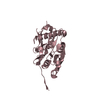
- PDB-7nwq: A carbohydrate binding module family 9 (CBM9) from Caldicellulosi... -
+ Open data

- Basic information
| Entry | Database: PDB / ID: 7nwq | |||||||||||||||
|---|---|---|---|---|---|---|---|---|---|---|---|---|---|---|---|---|
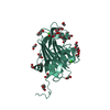
| Title | A carbohydrate binding module family 9 (CBM9) from Caldicellulosiruptor kristjanssonii in complex with cellotriose | |||||||||||||||
 Components Components | Beta-xylanase Xylanase Xylanase | |||||||||||||||
 Keywords Keywords | SUGAR BINDING PROTEIN / CARBOHYDRATE BINDING MODULE | |||||||||||||||
| Function / homology |  Function and homology informationendo-1,4-beta-xylanase activity / endo-1,4-beta-xylanase / polysaccharide catabolic process / carbohydrate binding Function and homology informationendo-1,4-beta-xylanase activity / endo-1,4-beta-xylanase / polysaccharide catabolic process / carbohydrate bindingSimilarity search - Function | |||||||||||||||
| Biological species | Caldicellulosiruptor kristjanssonii | |||||||||||||||
| Method | X-RAY DIFFRACTION / SYNCHROTRON / MOLECULAR REPLACEMENT / Resolution: 2.231 Å | |||||||||||||||
 Authors Authors | Krska, D. / Mazurkewich, S. / Navarro Poulsen, J. / Larsbrink, J. / Lo Leggio, L. | |||||||||||||||
| Funding support |  Sweden, Sweden,  Denmark, 4items Denmark, 4items
| |||||||||||||||
 Citation Citation | Journal: Biochemistry / Year: 2021 Title: Structural and Functional Analysis of a Multimodular Hyperthermostable Xylanase-Glucuronoyl Esterase from Caldicellulosiruptor kristjansonii . Authors: Krska, D. / Mazurkewich, S. / Brown, H.A. / Theibich, Y. / Poulsen, J.N. / Morris, A.L. / Koropatkin, N.M. / Lo Leggio, L. / Larsbrink, J. | |||||||||||||||
| History |
|
- Structure visualization
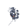
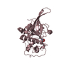
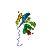
| Structure viewer | Molecule: MolmilJmol/JSmol |
|---|
- Downloads & links
-Download
| PDBx/mmCIF format | 7nwq.cif.gz | 66.8 KB | Display | PDBx/mmCIF format |
|---|---|---|---|---|
| PDB format | pdb7nwq.ent.gz | Display | PDB format | |
| PDBx/mmJSON format | 7nwq.json.gz | Tree view | PDBx/mmJSON format | |
| Others |  Other downloads Other downloads |
-Validation report
| Arichive directory | https://data.pdbj.org/pub/pdb/validation_reports/nw/7nwqftp://data.pdbj.org/pub/pdb/validation_reports/nw/7nwq | HTTPS FTP |
|---|
-Related structure data
| Related structure data | 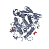 7nn3C  7nwnC 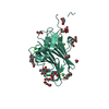 7nwoC 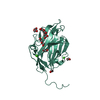 7nwpC 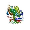 1i8uS S: Starting model for refinement C: citing same article ( |
|---|---|
| Similar structure data |


-Links
 PDBj
PDBj
- Assembly
| Deposited unit | 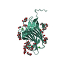
| |||||||||||||||
|---|---|---|---|---|---|---|---|---|---|---|---|---|---|---|---|---|
| 1 |
| |||||||||||||||
| Unit cell |
| |||||||||||||||
| Components on special symmetry positions |
|

-Components
-Protein / Sugars , 2 types, 2 molecules AAA
| #1: Protein | Xylanase Mass: 24645.232 Da / Num. of mol.: 1 Source method: isolated from a genetically manipulated source Source: (gene. exp.)  Caldicellulosiruptor kristjanssonii (strain ATCC 700853 / DSM 12137 / I77R1B) (bacteria) Caldicellulosiruptor kristjanssonii (strain ATCC 700853 / DSM 12137 / I77R1B) (bacteria)Strain: ATCC 700853 / DSM 12137 / I77R1B / Gene: Calkr_2245 / Production host: Escherichia coli BL21 (bacteria) / References: UniProt: E4S6E9, endo-1,4-beta-xylanase |
|---|---|
| #2: Polysaccharide | beta-D-glucopyranose-(1-4)-beta-D-glucopyranose-(1-4)-beta-D-glucopyranose  , Oligosaccharide / Class: Metabolism / Mass: 504.438 Da / Num. of mol.: 1 , Oligosaccharide / Class: Metabolism / Mass: 504.438 Da / Num. of mol.: 1Source method: isolated from a genetically manipulated source Details: oligosaccharide / References: beta-cellotriose |
-Non-polymers , 6 types, 133 molecules 


| #3: Chemical | ChemComp-PEG / Diethylene glycol Mass: 106.120 Da / Num. of mol.: 7 / Source method: obtained synthetically / Formula: C4H10O3 Mass: 106.120 Da / Num. of mol.: 7 / Source method: obtained synthetically / Formula: C4H10O3#4: Chemical | ChemComp-EDO / Ethylene glycol Mass: 62.068 Da / Num. of mol.: 9 / Source method: obtained synthetically / Formula: C2H6O2 Mass: 62.068 Da / Num. of mol.: 9 / Source method: obtained synthetically / Formula: C2H6O2#5: Chemical |  Mass: 40.078 Da / Num. of mol.: 3 / Source method: obtained synthetically / Formula: Ca Mass: 40.078 Da / Num. of mol.: 3 / Source method: obtained synthetically / Formula: Ca#6: Chemical | Polyethylene glycol Mass: 150.173 Da / Num. of mol.: 2 / Source method: obtained synthetically / Formula: C6H14O4 Mass: 150.173 Da / Num. of mol.: 2 / Source method: obtained synthetically / Formula: C6H14O4#7: Chemical | ChemComp-1PE / | Polyethylene glycol Mass: 238.278 Da / Num. of mol.: 1 / Source method: obtained synthetically / Formula: C10H22O6 / Comment: precipitant*YM Mass: 238.278 Da / Num. of mol.: 1 / Source method: obtained synthetically / Formula: C10H22O6 / Comment: precipitant*YM#8: Water | ChemComp-HOH / | WaterMass: 18.015 Da / Num. of mol.: 111 / Source method: isolated from a natural source / Formula: H2O |
|---|
-Details
| Has ligand of interest | Y |
|---|
-Experimental details
-Experiment
| Experiment | Method: X-RAY DIFFRACTION / Number of used crystals: 1 |
|---|
- Sample preparation
| Crystal | Density Matthews: 4.8 Å3/Da / Density % sol: 74.36 % |
|---|---|
| Crystal grow | Temperature: 293 K / Method: vapor diffusion, sitting drop / pH: 4.2 Details: 0.1 M phosphate-citrate and 40% polyethylene glycol 300 |
-Data collection
| Diffraction | Mean temperature: 100 K / Serial crystal experiment: N |
|---|---|
| Diffraction source | Source: SYNCHROTRON / Site: PETRA III, EMBL c/o DESY  / Beamline: P13 (MX1) / Wavelength: 1 Å / Beamline: P13 (MX1) / Wavelength: 1 Å |
| Detector | Type: DECTRIS PILATUS 6M-F / Detector: PIXEL / Date: Oct 5, 2019 |
| Radiation | Protocol: SINGLE WAVELENGTH / Monochromatic (M) / Laue (L): M / Scattering type: x-ray |
| Radiation wavelength | Wavelength: 1 Å / Relative weight: 1 |
| Reflection | Resolution: 2.23→70.52 Å / Num. obs: 21538 / % possible obs: 99.6 % / Redundancy: 71.8 % / CC1/2: 1 / Net I/σ(I): 26.89 |
| Reflection shell | Resolution: 2.23→2.29 Å / Num. unique obs: 1498 / CC1/2: 0.449 |
- Processing
| Software |
| |||||||||||||||||||||||||||||||||||||||||||||||||||||||||||||||||||||||||||||||||||||||||||||||||||||||||||||||||||||||||||||||||||||||||||||||||||
|---|---|---|---|---|---|---|---|---|---|---|---|---|---|---|---|---|---|---|---|---|---|---|---|---|---|---|---|---|---|---|---|---|---|---|---|---|---|---|---|---|---|---|---|---|---|---|---|---|---|---|---|---|---|---|---|---|---|---|---|---|---|---|---|---|---|---|---|---|---|---|---|---|---|---|---|---|---|---|---|---|---|---|---|---|---|---|---|---|---|---|---|---|---|---|---|---|---|---|---|---|---|---|---|---|---|---|---|---|---|---|---|---|---|---|---|---|---|---|---|---|---|---|---|---|---|---|---|---|---|---|---|---|---|---|---|---|---|---|---|---|---|---|---|---|---|---|---|---|
| Refinement | Method to determine structure: MOLECULAR REPLACEMENT Starting model: 1i8u Resolution: 2.231→70.518 Å / Cor.coef. Fo:Fc: 0.956 / Cor.coef. Fo:Fc free: 0.94 / WRfactor Rfree: 0.2 / WRfactor Rwork: 0.168 / SU B: 4.034 / SU ML: 0.097 / Average fsc free: 0.9448 / Average fsc overall: 0.956 / Average fsc work: 0.9572 / Cross valid method: FREE R-VALUE / ESU R: 0.169 / ESU R Free: 0.162 / Details: Hydrogens have not been used
| |||||||||||||||||||||||||||||||||||||||||||||||||||||||||||||||||||||||||||||||||||||||||||||||||||||||||||||||||||||||||||||||||||||||||||||||||||
| Solvent computation | Ion probe radii: 0.8 Å / Shrinkage radii: 0.8 Å / VDW probe radii: 1.2 Å / Solvent model: MASK BULK SOLVENT | |||||||||||||||||||||||||||||||||||||||||||||||||||||||||||||||||||||||||||||||||||||||||||||||||||||||||||||||||||||||||||||||||||||||||||||||||||
| Displacement parameters | Biso mean: 41.91 Å2
| |||||||||||||||||||||||||||||||||||||||||||||||||||||||||||||||||||||||||||||||||||||||||||||||||||||||||||||||||||||||||||||||||||||||||||||||||||
| Refinement step | Cycle: LAST / Resolution: 2.231→70.518 Å
| |||||||||||||||||||||||||||||||||||||||||||||||||||||||||||||||||||||||||||||||||||||||||||||||||||||||||||||||||||||||||||||||||||||||||||||||||||
| Refine LS restraints |
| |||||||||||||||||||||||||||||||||||||||||||||||||||||||||||||||||||||||||||||||||||||||||||||||||||||||||||||||||||||||||||||||||||||||||||||||||||
| LS refinement shell |
|
