 Movie
Movie Controller

[English] 日本語
 Yorodumi
- PDB-7nns: Crystal structure of the ACVR1 (ALK2) kinase in complex with the ... -
+ Open data

- Basic information
| Entry | Database: PDB / ID: 7nns | ||||||
|---|---|---|---|---|---|---|---|
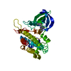
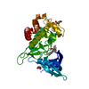
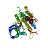
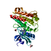
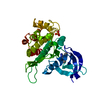
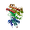
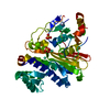
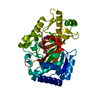
| Title | Crystal structure of the ACVR1 (ALK2) kinase in complex with the compound Momelotinib | ||||||
 Components Components | Activin receptor type I | ||||||
 Keywords Keywords |  SIGNALING PROTEIN / KINASE / BMP / INHIBITOR / SIGNALLING SIGNALING PROTEIN / KINASE / BMP / INHIBITOR / SIGNALLING | ||||||
| Function / homology |  Function and homology information Function and homology informationendocardial cushion cell fate commitment / mitral valve morphogenesis / endocardial cushion fusion / BMP receptor complex / atrial septum primum morphogenesis / BMP receptor activity / cardiac muscle cell fate commitment / acute inflammatory response / activin receptor activity, type I / positive regulation of cardiac epithelial to mesenchymal transition ...endocardial cushion cell fate commitment / mitral valve morphogenesis / endocardial cushion fusion / BMP receptor complex / atrial septum primum morphogenesis / BMP receptor activity / cardiac muscle cell fate commitment / acute inflammatory response / activin receptor activity, type I / positive regulation of cardiac epithelial to mesenchymal transition / positive regulation of determination of dorsal identity / transforming growth factor beta receptor activity, type I / activin receptor complex / endocardial cushion formation / smooth muscle cell differentiation / receptor protein serine/threonine kinase / transmembrane receptor protein serine/threonine kinase activity / pharyngeal system development / activin binding / cellular response to BMP stimulus / activin receptor signaling pathway / negative regulation of activin receptor signaling pathway / transforming growth factor beta binding / embryonic heart tube morphogenesis / gastrulation with mouth forming second / dorsal/ventral pattern formation / determination of left/right symmetry / neural crest cell migration / atrioventricular valve morphogenesis / branching involved in blood vessel morphogenesis / negative regulation of G1/S transition of mitotic cell cycle / ventricular septum morphogenesis / SMAD binding / germ cell development / positive regulation of SMAD protein signal transduction / peptide hormone binding / mesoderm formation / regulation of ossification / BMP signaling pathway / positive regulation of bone mineralization / positive regulation of osteoblast differentiation / negative regulation of signal transduction / transforming growth factor beta receptor signaling pathway / protein tyrosine kinase binding / negative regulation of extrinsic apoptotic signaling pathway / cellular response to growth factor stimulus / positive regulation of peptidyl-tyrosine phosphorylation / apical part of cell / heart development / in utero embryonic development / protein kinase activity / positive regulation of cell migration / cadherin binding / phosphorylation / protein serine/threonine kinase activity / positive regulation of DNA-templated transcription / protein homodimerization activity / positive regulation of transcription by RNA polymerase II / ATP binding / metal ion binding / plasma membraneSimilarity search - Function | ||||||
| Biological species |  Homo sapiens (human) Homo sapiens (human) | ||||||
| Method | X-RAY DIFFRACTION / SYNCHROTRON / MOLECULAR REPLACEMENT / Resolution: 2.14 Å | ||||||
 Authors Authors | Williams, E. / Chen, Z. / Burgess-Brown, N. / von Delft, F. / Arrowsmith, C.H. / Edwards, A.M. / Bountra, C. / Bullock, A.N. | ||||||
 Citation Citation | Journal: To Be Published Title: Crystal structure of the ACVR1 (ALK2) kinase in complex with the compound Momelotinib Authors: Williams, E. / Chen, Z. / Burgess-Brown, N. / von Delft, F. / Arrowsmith, C.H. / Edwards, A.M. / Bountra, C. / Bullock, A.N. | ||||||
| History |
|
- Structure visualization
| Structure viewer | Molecule: MolmilJmol/JSmol |
|---|
- Downloads & links
-Download
| PDBx/mmCIF format | 7nns.cif.gz | 80.6 KB | Display | PDBx/mmCIF format |
|---|---|---|---|---|
| PDB format | pdb7nns.ent.gz | 58.4 KB | Display | PDB format |
| PDBx/mmJSON format | 7nns.json.gz | Tree view | PDBx/mmJSON format | |
| Others |  Other downloads Other downloads |
-Validation report
| Arichive directory | https://data.pdbj.org/pub/pdb/validation_reports/nn/7nnsftp://data.pdbj.org/pub/pdb/validation_reports/nn/7nns | HTTPS FTP |
|---|
-Related structure data
| Related structure data |  3h9rS S: Starting model for refinement |
|---|---|
| Similar structure data |


-Links
 PDBj
PDBj
- Assembly
| Deposited unit | 
| ||||||||
|---|---|---|---|---|---|---|---|---|---|
| 1 |
| ||||||||
| Unit cell |
| ||||||||
| Components on special symmetry positions |
|
-Components
| #1: Protein | Mass: 34537.633 Da / Num. of mol.: 1 Source method: isolated from a genetically manipulated source Details: SEQUENCE CONTAINS A DELIBERATE MUTATION AT Q207D / Source: (gene. exp.) Homo sapiens (human) / Gene: ACVR1 / Cell line (production host): Sf9 / Production host:   Spodoptera frugiperda (fall armyworm) Spodoptera frugiperda (fall armyworm)References: UniProt: Q04771, receptor protein serine/threonine kinase | ||||||||
|---|---|---|---|---|---|---|---|---|---|
| #2: Chemical | ChemComp-EDO / Ethylene glycol  Mass: 62.068 Da / Num. of mol.: 6 / Source method: obtained synthetically / Formula: C2H6O2 Mass: 62.068 Da / Num. of mol.: 6 / Source method: obtained synthetically / Formula: C2H6O2#3: Chemical | ChemComp-C87 / | Momelotinib  Mass: 414.460 Da / Num. of mol.: 1 / Source method: obtained synthetically / Formula: C23H22N6O2 / Feature type: SUBJECT OF INVESTIGATION / Comment: medication, anticancer, inhibitor*YM Mass: 414.460 Da / Num. of mol.: 1 / Source method: obtained synthetically / Formula: C23H22N6O2 / Feature type: SUBJECT OF INVESTIGATION / Comment: medication, anticancer, inhibitor*YM#4: Chemical | ChemComp-SO4 / Sulfate  Mass: 96.063 Da / Num. of mol.: 7 / Source method: obtained synthetically / Formula: SO4 Mass: 96.063 Da / Num. of mol.: 7 / Source method: obtained synthetically / Formula: SO4#5: Water | ChemComp-HOH / | Water Mass: 18.015 Da / Num. of mol.: 116 / Source method: isolated from a natural source / Formula: H2O Mass: 18.015 Da / Num. of mol.: 116 / Source method: isolated from a natural source / Formula: H2OHas ligand of interest | Y | |
-Experimental details
-Experiment
| Experiment | Method: X-RAY DIFFRACTION / Number of used crystals: 1 |
|---|
- Sample preparation
| Crystal | Density Matthews: 2.71 Å3/Da / Density % sol: 54.63 % / Description: Single chain in the ASU |
|---|---|
| Crystal grow | Temperature: 293 K / Method: vapor diffusion, sitting drop / pH: 7.5 Details: 1.4M ammonium sulfate, 0.1M tris pH 7.5, 8% glycerol. |
-Data collection
| Diffraction | Mean temperature: 100 K / Serial crystal experiment: N |
|---|---|
| Diffraction source | Source: SYNCHROTRON / Site: Diamond  / Beamline: I04-1 / Wavelength: 0.92819 Å / Beamline: I04-1 / Wavelength: 0.92819 Å |
| Detector | Type: MARMOSAIC 300 mm CCD / Detector: CCD / Date: Jan 14, 2017 |
| Radiation | Monochromator: single bounce / Protocol: SINGLE WAVELENGTH / Monochromatic (M) / Laue (L): M / Scattering type: x-ray |
| Radiation wavelength | Wavelength: 0.92819 Å / Relative weight: 1 |
| Reflection | Resolution: 2.14→53.82 Å / Num. obs: 21731 / % possible obs: 100 % / Redundancy: 9.6 % / Biso Wilson estimate: 27.283 Å2 / CC1/2: 0.996 / Rmerge(I) obs: 0.24 / Rpim(I) all: 0.081 / Rrim(I) all: 0.253 / Net I/σ(I): 6.9 |
| Reflection shell | Resolution: 2.14→2.18 Å / Redundancy: 10 % / Rmerge(I) obs: 2.015 / Mean I/σ(I) obs: 1.1 / Num. unique obs: 1069 / CC1/2: 0.832 / Rpim(I) all: 0.668 / % possible all: 99.7 |
- Processing
| Software |
| ||||||||||||||||||||||||||||||||||||||||||||||||||||||||
|---|---|---|---|---|---|---|---|---|---|---|---|---|---|---|---|---|---|---|---|---|---|---|---|---|---|---|---|---|---|---|---|---|---|---|---|---|---|---|---|---|---|---|---|---|---|---|---|---|---|---|---|---|---|---|---|---|---|
| Refinement | Method to determine structure: MOLECULAR REPLACEMENT Starting model: 3H9R Resolution: 2.14→53.82 Å / SU ML: 0.36 / Cross valid method: THROUGHOUT / σ(F): 1.33 / Phase error: 31.96 / Stereochemistry target values: ML
| ||||||||||||||||||||||||||||||||||||||||||||||||||||||||
| Solvent computation | Shrinkage radii: 0.9 Å / VDW probe radii: 1.11 Å / Solvent model: FLAT BULK SOLVENT MODEL | ||||||||||||||||||||||||||||||||||||||||||||||||||||||||
| Displacement parameters | Biso max: 79.23 Å2 / Biso mean: 28.5963 Å2 / Biso min: 11.24 Å2 | ||||||||||||||||||||||||||||||||||||||||||||||||||||||||
| Refinement step | Cycle: final / Resolution: 2.14→53.82 Å
| ||||||||||||||||||||||||||||||||||||||||||||||||||||||||
| LS refinement shell | Refine-ID: X-RAY DIFFRACTION / Rfactor Rfree error: 0 / Total num. of bins used: 7
|
