 Movie
Movie Controller

[English] 日本語
 Yorodumi
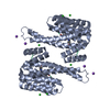
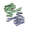
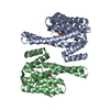
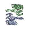
- PDB-7nmz: Structure of 14-3-3 eta in complex with Nedd4-2(335-455) containi... -
+ Open data

- Basic information
| Entry | Database: PDB / ID: 7nmz | ||||||
|---|---|---|---|---|---|---|---|
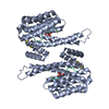
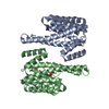
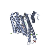
| Title | Structure of 14-3-3 eta in complex with Nedd4-2(335-455) containing two 14-3-3 binding motifs Ser342 and Ser448 | ||||||
 Components Components |
| ||||||
 Keywords Keywords |  SIGNALING PROTEIN / E3 ubiquitin protein ligase / complex / Nedd4-2 / 14-3-3 protein SIGNALING PROTEIN / E3 ubiquitin protein ligase / complex / Nedd4-2 / 14-3-3 protein | ||||||
| Function / homology |  Function and homology information Function and homology informationglucocorticoid catabolic process / cerebellar granule cell to Purkinje cell synapse / positive regulation of caveolin-mediated endocytosis / RING-type E3 ubiquitin transferase (cysteine targeting) / negative regulation of sodium ion transmembrane transport / negative regulation of potassium ion transmembrane transporter activity / negative regulation of dendrite morphogenesis / regulation of potassium ion transmembrane transporter activity / negative regulation of potassium ion transmembrane transport / intracellular glucocorticoid receptor signaling pathway ...glucocorticoid catabolic process / cerebellar granule cell to Purkinje cell synapse / positive regulation of caveolin-mediated endocytosis / RING-type E3 ubiquitin transferase (cysteine targeting) / negative regulation of sodium ion transmembrane transport / negative regulation of potassium ion transmembrane transporter activity / negative regulation of dendrite morphogenesis / regulation of potassium ion transmembrane transporter activity / negative regulation of potassium ion transmembrane transport / intracellular glucocorticoid receptor signaling pathway / regulation of sodium ion transmembrane transport / negative regulation of sodium ion transmembrane transporter activity / regulation of sodium ion transmembrane transporter activity / negative regulation of protein localization to cell surface / regulation of membrane repolarization / nuclear glucocorticoid receptor binding / positive regulation of dendrite extension / potassium channel inhibitor activity / ventricular cardiac muscle cell action potential / HECT-type E3 ubiquitin transferase / membrane depolarization during action potential / sodium channel inhibitor activity / regulation of monoatomic ion transmembrane transport / regulation of neuron differentiation / regulation of dendrite morphogenesis / regulation of membrane depolarization / protein monoubiquitination / intercalated disc / sodium channel regulator activity / Activation of BAD and translocation to mitochondria / potassium channel regulator activity / protein K48-linked ubiquitination / SARS-CoV-2 targets host intracellular signalling and regulatory pathways / Chk1/Chk2(Cds1) mediated inactivation of Cyclin B:Cdk1 complex / SARS-CoV-1 targets host intracellular signalling and regulatory pathways / regulation of sodium ion transport / RHO GTPases activate PKNs / monoatomic ion transmembrane transport / insulin-like growth factor receptor binding / substantia nigra development / presynaptic modulation of chemical synaptic transmission / multivesicular body / Downregulation of TGF-beta receptor signaling / regulation of membrane potential / Translocation of SLC2A4 (GLUT4) to the plasma membrane / TP53 Regulates Metabolic Genes / Downregulation of SMAD2/3:SMAD4 transcriptional activity / intracellular protein transport / regulation of synaptic plasticity / Budding and maturation of HIV virion / regulation of protein stability / Stimuli-sensing channels / positive regulation of protein catabolic process / ubiquitin-protein transferase activity / ubiquitin protein ligase activity / Antigen processing: Ubiquitination & Proteasome degradation / presynapse / actin binding / ubiquitin-dependent protein catabolic process / proteasome-mediated ubiquitin-dependent protein catabolic process / transmembrane transporter binding / cell differentiation / protein ubiquitination / protein heterodimerization activity / protein domain specific binding / positive regulation of DNA-templated transcription / Golgi apparatus / enzyme binding / signal transduction / extracellular exosome / nucleoplasm / identical protein binding / plasma membrane / cytosol / cytoplasmSimilarity search - Function | ||||||
| Biological species |  Homo sapiens (human) Homo sapiens (human) | ||||||
| Method | X-RAY DIFFRACTION / MOLECULAR REPLACEMENT / Resolution: 2.303 Å | ||||||
 Authors Authors | Pohl, P. / Obsil, T. / Obsilova, V. | ||||||
| Funding support |  Czech Republic, 1items Czech Republic, 1items
| ||||||
 Citation Citation | Journal: Commun Biol / Year: 2021 Title: 14-3-3-protein regulates Nedd4-2 by modulating interactions between HECT and WW domains. Authors: Pohl, P. / Joshi, R. / Petrvalska, O. / Obsil, T. / Obsilova, V. | ||||||
| History |
|
- Structure visualization
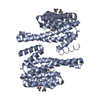
| Structure viewer | Molecule: MolmilJmol/JSmol |
|---|
- Downloads & links
-Download
| PDBx/mmCIF format | 7nmz.cif.gz | 134.5 KB | Display | PDBx/mmCIF format |
|---|---|---|---|---|
| PDB format | pdb7nmz.ent.gz | Display | PDB format | |
| PDBx/mmJSON format | 7nmz.json.gz | Tree view | PDBx/mmJSON format | |
| Others |  Other downloads Other downloads |
-Validation report
| Arichive directory | https://data.pdbj.org/pub/pdb/validation_reports/nm/7nmzftp://data.pdbj.org/pub/pdb/validation_reports/nm/7nmz | HTTPS FTP |
|---|
-Related structure data
| Related structure data |  6zbtC  6zc9C  2c63S S: Starting model for refinement C: citing same article ( |
|---|---|
| Similar structure data |

-Links
 PDBj
PDBj


- Assembly
| Deposited unit | 
| ||||||||||||
|---|---|---|---|---|---|---|---|---|---|---|---|---|---|
| 1 |
| ||||||||||||
| Unit cell |
|
-Components
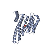
| #1: Protein | Mass: 27186.777 Da / Num. of mol.: 2 / Mutation: S235Stop Source method: isolated from a genetically manipulated source Source: (gene. exp.) Homo sapiens (human) / Gene: YWHAH, YWHA1 / Production host:  Escherichia coli BL21(DE3) (bacteria) / References: UniProt: Q04917 Escherichia coli BL21(DE3) (bacteria) / References: UniProt: Q04917#2: Protein | | Mass: 13442.557 Da / Num. of mol.: 1 / Mutation: T367A Source method: isolated from a genetically manipulated source Source: (gene. exp.) Homo sapiens (human) / Gene: NEDD4L, KIAA0439, NEDL3 / Production host: Escherichia coli BL21(DE3) (bacteria)References: UniProt: Q96PU5, HECT-type E3 ubiquitin transferase #3: Water | ChemComp-HOH / | Water Mass: 18.015 Da / Num. of mol.: 189 / Source method: isolated from a natural source / Formula: H2O Mass: 18.015 Da / Num. of mol.: 189 / Source method: isolated from a natural source / Formula: H2OHas ligand of interest | N | |
|---|
-Experimental details
-Experiment
| Experiment | Method: X-RAY DIFFRACTION / Number of used crystals: 1 |
|---|
- Sample preparation
| Crystal | Density Matthews: 2.32 Å3/Da / Density % sol: 47 % |
|---|---|
| Crystal grow | Temperature: 293.15 K / Method: vapor diffusion, hanging drop / pH: 7.5 Details: 12.5% w/v PEG 1000, 12.5% w/v PEG 3350, 12.5% v/v MPD, 0.03M of each NPS (sodium nitrate, sodium phosphate dibasic, ammonium sulfate), 0.1Mbicine/Trizma base pH 8.5, 30% sacharose |
-Data collection
| Diffraction | Mean temperature: 100 K / Serial crystal experiment: N |
|---|---|
| Diffraction source | Source: LIQUID ANODE / Type: Excillum MetalJet D2+ 70 kV / Wavelength: 1.3418 Å |
| Detector | Type: Bruker PHOTON II / Detector: PIXEL / Date: Jan 20, 2021 |
| Radiation | Protocol: SINGLE WAVELENGTH / Monochromatic (M) / Laue (L): M / Scattering type: x-ray |
| Radiation wavelength | Wavelength: 1.3418 Å / Relative weight: 1 |
| Reflection | Resolution: 2.303→32.69 Å / Num. obs: 32703 / % possible obs: 99.86 % / Redundancy: 4.73 % / Biso Wilson estimate: 33.19 Å2 / Rrim(I) all: 0.064 / Net I/σ(I): 19.68 |
| Reflection shell | Resolution: 2.303→2.385 Å / Redundancy: 3.99 % / Mean I/σ(I) obs: 1.94 / Num. unique obs: 3215 / Rrim(I) all: 0.655 |
- Processing
| Software |
| |||||||||||||||||||||||||||||||||||||||||||||||||||||||||||||||||||||||||||||||||||||||||||
|---|---|---|---|---|---|---|---|---|---|---|---|---|---|---|---|---|---|---|---|---|---|---|---|---|---|---|---|---|---|---|---|---|---|---|---|---|---|---|---|---|---|---|---|---|---|---|---|---|---|---|---|---|---|---|---|---|---|---|---|---|---|---|---|---|---|---|---|---|---|---|---|---|---|---|---|---|---|---|---|---|---|---|---|---|---|---|---|---|---|---|---|---|
| Refinement | Method to determine structure: MOLECULAR REPLACEMENT Starting model: 2C63 Resolution: 2.303→32.69 Å / SU ML: 0.1987 / Cross valid method: FREE R-VALUE / σ(F): 1.36 / Phase error: 24.5299 Stereochemistry target values: GeoStd + Monomer Library + CDL v1.2
| |||||||||||||||||||||||||||||||||||||||||||||||||||||||||||||||||||||||||||||||||||||||||||
| Solvent computation | Shrinkage radii: 0.9 Å / VDW probe radii: 1.11 Å / Solvent model: FLAT BULK SOLVENT MODEL | |||||||||||||||||||||||||||||||||||||||||||||||||||||||||||||||||||||||||||||||||||||||||||
| Displacement parameters | Biso mean: 43.92 Å2 | |||||||||||||||||||||||||||||||||||||||||||||||||||||||||||||||||||||||||||||||||||||||||||
| Refinement step | Cycle: LAST / Resolution: 2.303→32.69 Å
| |||||||||||||||||||||||||||||||||||||||||||||||||||||||||||||||||||||||||||||||||||||||||||
| Refine LS restraints |
| |||||||||||||||||||||||||||||||||||||||||||||||||||||||||||||||||||||||||||||||||||||||||||
| LS refinement shell |
|
