 Movie
Movie Controller

[English] 日本語
 Yorodumi
- PDB-7njz: X-ray crystallography study of RoAb13 which binds to PIYDIN, a pa... -
+ Open data

- Basic information
| Entry | Database: PDB / ID: 7njz | ||||||
|---|---|---|---|---|---|---|---|
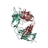
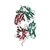
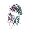
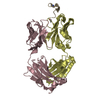
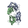
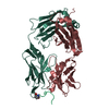
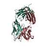
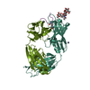
| Title | X-ray crystallography study of RoAb13 which binds to PIYDIN, a part of the CCR5 N terminal domain | ||||||
 Components Components |
| ||||||
 Keywords Keywords |  IMMUNE SYSTEM / CCR5 Peptidomimetic IMMUNE SYSTEM / CCR5 Peptidomimetic | ||||||
| Function / homology |  Function and homology information Function and homology informationchemokine (C-C motif) ligand 5 binding / negative regulation of macrophage apoptotic process / signaling / chemokine receptor activity / C-C chemokine receptor activity / C-C chemokine binding / phosphatidylinositol phospholipase C activity / response to cholesterol / Chemokine receptors bind chemokines / release of sequestered calcium ion into cytosol by sarcoplasmic reticulum ...chemokine (C-C motif) ligand 5 binding / negative regulation of macrophage apoptotic process / signaling / chemokine receptor activity / C-C chemokine receptor activity / C-C chemokine binding / phosphatidylinositol phospholipase C activity / response to cholesterol / Chemokine receptors bind chemokines / release of sequestered calcium ion into cytosol by sarcoplasmic reticulum / dendritic cell chemotaxis / Interleukin-10 signaling / Binding and entry of HIV virion / cellular defense response / coreceptor activity / cell chemotaxis / calcium-mediated signaling / calcium ion transport / chemotaxis / MAPK cascade / virus receptor activity / cell-cell signaling / actin binding / G alpha (i) signalling events / positive regulation of cytosolic calcium ion concentration / cellular response to lipopolysaccharide / cell surface receptor signaling pathway / endosome / inflammatory response / immune response / G protein-coupled receptor signaling pathway / external side of plasma membrane / cell surface / identical protein binding / plasma membrane / cytoplasmSimilarity search - Function | ||||||
| Biological species |  Mus musculus (house mouse) Mus musculus (house mouse) Homo sapiens (human) Homo sapiens (human) | ||||||
| Method | X-RAY DIFFRACTION / SYNCHROTRON / MOLECULAR REPLACEMENT / Resolution: 3.2 Å | ||||||
 Authors Authors | Helliwell, J.R. / Chayen, N. / Saridakis, E. / Govada, L. | ||||||
| Funding support |  United Kingdom, 1items United Kingdom, 1items
| ||||||
 Citation Citation | Journal: Iucrj / Year: 2021 Title: X-ray crystallographic studies of RoAb13 bound to PIYDIN, a part of the N-terminal domain of C-C chemokine receptor 5. Authors: Govada, L. / Saridakis, E. / Kassen, S.C. / Bin-Ramzi, A. / Morgan, R.M. / Chain, B. / Helliwell, J.R. / Chayen, N.E. | ||||||
| History |
|
- Structure visualization
| Structure viewer | Molecule: MolmilJmol/JSmol |
|---|
- Downloads & links
-Download
| PDBx/mmCIF format | 7njz.cif.gz | 118 KB | Display | PDBx/mmCIF format |
|---|---|---|---|---|
| PDB format | pdb7njz.ent.gz | 74.3 KB | Display | PDB format |
| PDBx/mmJSON format | 7njz.json.gz | Tree view | PDBx/mmJSON format | |
| Others |  Other downloads Other downloads |
-Validation report
| Arichive directory | https://data.pdbj.org/pub/pdb/validation_reports/nj/7njzftp://data.pdbj.org/pub/pdb/validation_reports/nj/7njz | HTTPS FTP |
|---|
-Related structure data
| Related structure data | 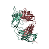 7nw3C  4s2sS S: Starting model for refinement C: citing same article ( |
|---|---|
| Similar structure data | |
| Experimental dataset #1 | Data reference: 10.5281/zenodo.4912886 / Data set type: diffraction image data |


-Links
 PDBj
PDBj


- Assembly
| Deposited unit | 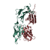
| ||||||||||||
|---|---|---|---|---|---|---|---|---|---|---|---|---|---|
| 1 | 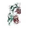
| ||||||||||||
| 2 |
| ||||||||||||
| Unit cell |
|
-Components
| #1: Antibody | Mass: 24063.961 Da / Num. of mol.: 1 / Source method: isolated from a natural source / Source: (natural) Mus musculus (house mouse) |
|---|---|
| #2: Antibody | Mass: 24459.154 Da / Num. of mol.: 1 / Source method: isolated from a natural source / Source: (natural) Mus musculus (house mouse) |
| #3: Protein/peptide | / C-C CKR-5 / CC-CKR-5 / CCR-5 / CCR5 / CHEMR13 / HIV-1 fusion coreceptor / PIYDIN Mass: 733.810 Da / Num. of mol.: 1 / Source method: obtained synthetically / Source: (synth.) Homo sapiens (human) / References: UniProt: P51681 |
| #4: Water | ChemComp-HOH / Water Mass: 18.015 Da / Num. of mol.: 22 / Source method: isolated from a natural source / Formula: H2O Mass: 18.015 Da / Num. of mol.: 22 / Source method: isolated from a natural source / Formula: H2O |
-Experimental details
-Experiment
| Experiment | Method: X-RAY DIFFRACTION / Number of used crystals: 1 |
|---|
- Sample preparation
| Crystal | Density Matthews: 3.88 Å3/Da / Density % sol: 68 % / Description: Bipyramidal. |
|---|---|
| Crystal grow | Temperature: 293 K / Method: vapor diffusion, hanging drop / Details: 1.85 M Ammonium sulfate / PH range: 5.0 - 6.0 Temp details: All experiments were incubated in a temperature controlled Incubator at 293K |
-Data collection
| Diffraction | Mean temperature: 100 K / Serial crystal experiment: N |
|---|---|
| Diffraction source | Source: SYNCHROTRON / Site: Diamond / Beamline: I04 / Wavelength: 0.97949 Å |
| Detector | Type: DECTRIS PILATUS 6M-F / Detector: PIXEL / Date: May 20, 2016 |
| Radiation | Protocol: SINGLE WAVELENGTH / Monochromatic (M) / Laue (L): M / Scattering type: x-ray |
| Radiation wavelength | Wavelength: 0.97949 Å / Relative weight: 1 |
| Reflection | Resolution: 3.2→73.53 Å / Num. obs: 13940 / % possible obs: 100 % / Redundancy: 7 % / Biso Wilson estimate: 121.9 Å2 / Rmerge(I) obs: 0.08 / Net I/σ(I): 7.6 |
| Reflection shell | Resolution: 3.2→3.5 Å / Rmerge(I) obs: 0.431 / Mean I/σ(I) obs: 3.2 / Num. unique obs: 2703 / CC1/2: 0.95 / % possible all: 100 |
- Processing
| Software |
| ||||||||||||||||||||||||||||||||||||||||||
|---|---|---|---|---|---|---|---|---|---|---|---|---|---|---|---|---|---|---|---|---|---|---|---|---|---|---|---|---|---|---|---|---|---|---|---|---|---|---|---|---|---|---|---|
| Refinement | Method to determine structure: MOLECULAR REPLACEMENT Starting model: 4S2S Resolution: 3.2→73.52 Å / SU ML: 0.6142 / Cross valid method: FREE R-VALUE / σ(F): 1.36 / Phase error: 33.1435 Stereochemistry target values: GeoStd + Monomer Library + CDL v1.2
| ||||||||||||||||||||||||||||||||||||||||||
| Solvent computation | Shrinkage radii: 0.9 Å / VDW probe radii: 1.11 Å / Solvent model: FLAT BULK SOLVENT MODEL | ||||||||||||||||||||||||||||||||||||||||||
| Displacement parameters | Biso mean: 121.77 Å2 | ||||||||||||||||||||||||||||||||||||||||||
| Refinement step | Cycle: LAST / Resolution: 3.2→73.52 Å
| ||||||||||||||||||||||||||||||||||||||||||
| Refine LS restraints |
| ||||||||||||||||||||||||||||||||||||||||||
| LS refinement shell |
|
