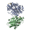
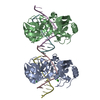
 Movie
Movie Controller


+ Open data

- Basic information
| Entry | Database: PDB / ID: 7nix | |||||||||
|---|---|---|---|---|---|---|---|---|---|---|
| Title | 14-3-3 sigma with AS160 binding site pT642 | |||||||||
 Components Components |
| |||||||||
 Keywords Keywords | PEPTIDE BINDING PROTEIN / 1433 / TBC1 domain family member 4 / peptide-protein complex | |||||||||
| Function / homology |  Function and homology information Function and homology informationnegative regulation of vesicle fusion / regulation of epidermal cell division /  protein kinase C inhibitor activity / positive regulation of epidermal cell differentiation / keratinocyte development / keratinization / Regulation of localization of FOXO transcription factors / keratinocyte proliferation / phosphoserine residue binding / Activation of BAD and translocation to mitochondria ...negative regulation of vesicle fusion / regulation of epidermal cell division / protein kinase C inhibitor activity / positive regulation of epidermal cell differentiation / keratinocyte development / keratinization / Regulation of localization of FOXO transcription factors / keratinocyte proliferation / phosphoserine residue binding / Activation of BAD and translocation to mitochondria / negative regulation of keratinocyte proliferation / establishment of skin barrier / SARS-CoV-2 targets host intracellular signalling and regulatory pathways / Chk1/Chk2(Cds1) mediated inactivation of Cyclin B:Cdk1 complex / protein kinase A signaling / negative regulation of stem cell proliferation / SARS-CoV-1 targets host intracellular signalling and regulatory pathways / RHO GTPases activate PKNs / vesicle-mediated transport / protein export from nucleus / negative regulation of innate immune response / GTPase activator activity / protein sequestering activity / TP53 Regulates Transcription of Genes Involved in G2 Cell Cycle Arrest / release of cytochrome c from mitochondria / positive regulation of protein export from nucleus / stem cell proliferation / Translocation of SLC2A4 (GLUT4) to the plasma membrane / TP53 Regulates Metabolic Genes / negative regulation of protein kinase activity / negative regulation of cysteine-type endopeptidase activity involved in apoptotic process / cellular response to insulin stimulus / intrinsic apoptotic signaling pathway in response to DNA damage / positive regulation of cell growth / vesicle / regulation of cell cycle / cadherin binding / protein kinase binding / negative regulation of transcription by RNA polymerase II / signal transduction / extracellular space / extracellular exosome / identical protein binding / nucleus / cytosol / cytoplasm protein kinase C inhibitor activity / positive regulation of epidermal cell differentiation / keratinocyte development / keratinization / Regulation of localization of FOXO transcription factors / keratinocyte proliferation / phosphoserine residue binding / Activation of BAD and translocation to mitochondria ...negative regulation of vesicle fusion / regulation of epidermal cell division / protein kinase C inhibitor activity / positive regulation of epidermal cell differentiation / keratinocyte development / keratinization / Regulation of localization of FOXO transcription factors / keratinocyte proliferation / phosphoserine residue binding / Activation of BAD and translocation to mitochondria / negative regulation of keratinocyte proliferation / establishment of skin barrier / SARS-CoV-2 targets host intracellular signalling and regulatory pathways / Chk1/Chk2(Cds1) mediated inactivation of Cyclin B:Cdk1 complex / protein kinase A signaling / negative regulation of stem cell proliferation / SARS-CoV-1 targets host intracellular signalling and regulatory pathways / RHO GTPases activate PKNs / vesicle-mediated transport / protein export from nucleus / negative regulation of innate immune response / GTPase activator activity / protein sequestering activity / TP53 Regulates Transcription of Genes Involved in G2 Cell Cycle Arrest / release of cytochrome c from mitochondria / positive regulation of protein export from nucleus / stem cell proliferation / Translocation of SLC2A4 (GLUT4) to the plasma membrane / TP53 Regulates Metabolic Genes / negative regulation of protein kinase activity / negative regulation of cysteine-type endopeptidase activity involved in apoptotic process / cellular response to insulin stimulus / intrinsic apoptotic signaling pathway in response to DNA damage / positive regulation of cell growth / vesicle / regulation of cell cycle / cadherin binding / protein kinase binding / negative regulation of transcription by RNA polymerase II / signal transduction / extracellular space / extracellular exosome / identical protein binding / nucleus / cytosol / cytoplasmSimilarity search - Function | |||||||||
| Biological species |  Homo sapiens (human) Homo sapiens (human) | |||||||||
| Method | X-RAY DIFFRACTION / SYNCHROTRON / MOLECULAR REPLACEMENT / molecular replacement / Resolution: 1.9 Å | |||||||||
 Authors Authors | Wolter, M. / Ottmann, C. | |||||||||
| Funding support | 2items
| |||||||||
 Citation Citation | Journal: J.Am.Chem.Soc. / Year: 2021 Title: Reversible Covalent Imine-Tethering for Selective Stabilization of 14-3-3 Hub Protein Interactions. Authors: Cossar, P.J. / Wolter, M. / van Dijck, L. / Valenti, D. / Levy, L.M. / Ottmann, C. / Brunsveld, L. | |||||||||
| History |
|
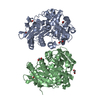
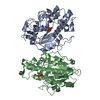
- Structure visualization
| Structure viewer | Molecule: MolmilJmol/JSmol |
|---|
- Downloads & links
-Download
| PDBx/mmCIF format | 7nix.cif.gz | 109.8 KB | Display | PDBx/mmCIF format |
|---|---|---|---|---|
| PDB format | pdb7nix.ent.gz | 83.7 KB | Display | PDB format |
| PDBx/mmJSON format | 7nix.json.gz | Tree view | PDBx/mmJSON format | |
| Others |  Other downloads Other downloads |
-Validation report
| Arichive directory | https://data.pdbj.org/pub/pdb/validation_reports/ni/7nixftp://data.pdbj.org/pub/pdb/validation_reports/ni/7nix | HTTPS FTP |
|---|
-Related structure data
| Related structure data | 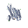 7aogC 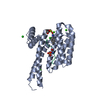 7axnC 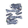 7ayfC 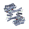 7az1C 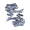 7az2C 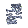 7bdpC 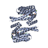 7bdtC 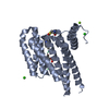 7bdyC 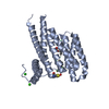 7bfwC 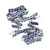 7bg3C 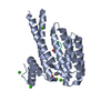 7bgqC 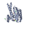 7bgrC 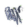 7bgvC 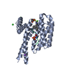 7bgwC 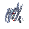 7nifC 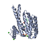 7nigC 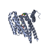 7nj6C 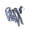 7nj8C 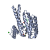 7njaC 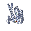 7nqpC 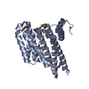 7nrkC 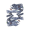 7nrlC 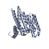 7nsvC 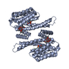 4jddS S: Starting model for refinement C: citing same article ( |
|---|---|
| Similar structure data |


-Links
 PDBj
PDBj


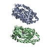
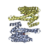
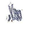
- Assembly
| Deposited unit | 
| |||||||||
|---|---|---|---|---|---|---|---|---|---|---|
| 1 | 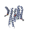
| |||||||||
| Unit cell |
| |||||||||
| Components on special symmetry positions |
|
-Components
| #1: Protein | Mass: 28210.518 Da / Num. of mol.: 1 Source method: isolated from a genetically manipulated source Source: (gene. exp.) Homo sapiens (human) / Gene: SFN, HME1 / Production host:  Escherichia coli (E. coli) / References: UniProt: P31947 Escherichia coli (E. coli) / References: UniProt: P31947 | ||||||
|---|---|---|---|---|---|---|---|
| #2: Protein/peptide | Mass: 1446.533 Da / Num. of mol.: 1 / Source method: obtained synthetically / Source: (synth.) Homo sapiens (human) / References: UniProt: O60343 | ||||||
| #3: Chemical | CHES (buffer)  Mass: 207.290 Da / Num. of mol.: 2 / Source method: obtained synthetically / Formula: C8H17NO3S / Comment: pH buffer*YM Mass: 207.290 Da / Num. of mol.: 2 / Source method: obtained synthetically / Formula: C8H17NO3S / Comment: pH buffer*YM#4: Chemical |   Mass: 24.305 Da / Num. of mol.: 2 / Source method: obtained synthetically / Formula: Mg Mass: 24.305 Da / Num. of mol.: 2 / Source method: obtained synthetically / Formula: Mg#5: Water | ChemComp-HOH / | Water Mass: 18.015 Da / Num. of mol.: 197 / Source method: isolated from a natural source / Formula: H2O Mass: 18.015 Da / Num. of mol.: 197 / Source method: isolated from a natural source / Formula: H2OHas ligand of interest | N | |
-Experimental details
-Experiment
| Experiment | Method: X-RAY DIFFRACTION / Number of used crystals: 1 |
|---|
- Sample preparation
| Crystal | Density Matthews: 2.66 Å3/Da / Density % sol: 53.8 % |
|---|---|
| Crystal grow | Temperature: 277 K / Method: vapor diffusion, sitting drop / pH: 9.5 Details: 40% (v/v) MPD, 100 mM CHES/ Sodium hydroxide pH 9.5 |
-Data collection
| Diffraction | Mean temperature: 100 K / Serial crystal experiment: N |
|---|---|
| Diffraction source | Source: SYNCHROTRON / Site: Diamond  / Beamline: I03 / Wavelength: 0.976284 Å / Beamline: I03 / Wavelength: 0.976284 Å |
| Detector | Type: DECTRIS EIGER2 XE 16M / Detector: PIXEL / Date: Feb 28, 2020 |
| Radiation | Protocol: SINGLE WAVELENGTH / Monochromatic (M) / Laue (L): M / Scattering type: x-ray |
| Radiation wavelength | Wavelength: 0.976284 Å / Relative weight: 1 |
| Reflection | Resolution: 1.9→104.98 Å / Num. obs: 25949 / % possible obs: 100 % / Redundancy: 37.5 % / Biso Wilson estimate: 29.31 Å2 / CC1/2: 1 / Rmerge(I) obs: 0.104 / Rpim(I) all: 0.017 / Rrim(I) all: 0.105 / Net I/σ(I): 25.4 |
| Reflection shell | Resolution: 1.9→1.94 Å / Redundancy: 38.7 % / Rmerge(I) obs: 1.55 / Num. unique obs: 1623 / CC1/2: 0.97 / Rpim(I) all: 0.252 / Rrim(I) all: 1.57 / % possible all: 100 |
-Phasing
| Phasing | Method: molecular replacement |
|---|
- Processing
| Software |
| ||||||||||||||||||||||||||||||||||||||||||||||||||||||||||||||||||||||||||||||||||||||||||||||||||||||||||||
|---|---|---|---|---|---|---|---|---|---|---|---|---|---|---|---|---|---|---|---|---|---|---|---|---|---|---|---|---|---|---|---|---|---|---|---|---|---|---|---|---|---|---|---|---|---|---|---|---|---|---|---|---|---|---|---|---|---|---|---|---|---|---|---|---|---|---|---|---|---|---|---|---|---|---|---|---|---|---|---|---|---|---|---|---|---|---|---|---|---|---|---|---|---|---|---|---|---|---|---|---|---|---|---|---|---|---|---|---|---|
| Refinement | Method to determine structure: MOLECULAR REPLACEMENT Starting model: 4JDD Resolution: 1.9→60.61 Å / SU ML: 0.23 / Cross valid method: FREE R-VALUE / σ(F): 1.34 / Phase error: 22.44 / Stereochemistry target values: ML
| ||||||||||||||||||||||||||||||||||||||||||||||||||||||||||||||||||||||||||||||||||||||||||||||||||||||||||||
| Solvent computation | Shrinkage radii: 0.9 Å / VDW probe radii: 1.11 Å / Solvent model: FLAT BULK SOLVENT MODEL | ||||||||||||||||||||||||||||||||||||||||||||||||||||||||||||||||||||||||||||||||||||||||||||||||||||||||||||
| Displacement parameters | Biso max: 115.06 Å2 / Biso mean: 37.2096 Å2 / Biso min: 15.24 Å2 | ||||||||||||||||||||||||||||||||||||||||||||||||||||||||||||||||||||||||||||||||||||||||||||||||||||||||||||
| Refinement step | Cycle: final / Resolution: 1.9→60.61 Å
| ||||||||||||||||||||||||||||||||||||||||||||||||||||||||||||||||||||||||||||||||||||||||||||||||||||||||||||
| LS refinement shell | Refine-ID: X-RAY DIFFRACTION / Rfactor Rfree error: 0 / Total num. of bins used: 17 / % reflection obs: 100 %
|
