 Movie
Movie Controller


+ Open data

- Basic information
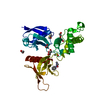
| Entry | Database: PDB / ID: 7ncf | ||||||
|---|---|---|---|---|---|---|---|
| Title | Crystal structure of HIPK2 in complex with MU135 (compound 21e) | ||||||
 Components Components | Homeodomain-interacting protein kinase 2 | ||||||
 Keywords Keywords |  TRANSFERASE / kinase / HIPK2 / kinase inhibitor / Structural Genomics / Structural Genomics Consortium / SGC TRANSFERASE / kinase / HIPK2 / kinase inhibitor / Structural Genomics / Structural Genomics Consortium / SGC | ||||||
| Function / homology |  Function and homology information Function and homology informationlens induction in camera-type eye / iris morphogenesis / embryonic retina morphogenesis in camera-type eye / embryonic camera-type eye morphogenesis / retina layer formation / Physiological factors / PML body organization / voluntary musculoskeletal movement / eye development / YAP1- and WWTR1 (TAZ)-stimulated gene expression ...lens induction in camera-type eye / iris morphogenesis / embryonic retina morphogenesis in camera-type eye / embryonic camera-type eye morphogenesis / retina layer formation / Physiological factors / PML body organization / voluntary musculoskeletal movement / eye development / YAP1- and WWTR1 (TAZ)-stimulated gene expression / SMAD protein signal transduction / adult walking behavior / virion binding / anterior/posterior pattern specification / DNA damage response, signal transduction by p53 class mediator resulting in transcription of p21 class mediator / smoothened signaling pathway / RUNX1 interacts with co-factors whose precise effect on RUNX1 targets is not known / positive regulation of transforming growth factor beta receptor signaling pathway / SMAD binding / Regulation of MECP2 expression and activity / negative regulation of BMP signaling pathway / intrinsic apoptotic signaling pathway in response to DNA damage by p53 class mediator / positive regulation of DNA binding / negative regulation of ubiquitin-dependent protein catabolic process / intrinsic apoptotic signaling pathway / erythrocyte differentiation / transforming growth factor beta receptor signaling pathway / SUMOylation of transcription cofactors / regulation of signal transduction by p53 class mediator / positive regulation of JNK cascade / peptidyl-threonine phosphorylation / neuron differentiation / PML body / positive regulation of DNA-binding transcription factor activity / cytoplasmic stress granule / RNA polymerase II transcription regulator complex / positive regulation of angiogenesis / transcription corepressor activity / positive regulation of protein binding / cellular response to hypoxia / peptidyl-serine phosphorylation / protein tyrosine kinase activity / neuron apoptotic process / Regulation of TP53 Activity through Phosphorylation / RNA polymerase II-specific DNA-binding transcription factor binding / negative regulation of neuron apoptotic process / cell population proliferation / transcription coactivator activity / nuclear body / non-specific serine/threonine protein kinase / regulation of cell cycle / protein kinase activity / positive regulation of protein phosphorylation / protein phosphorylation / protein serine kinase activity / protein serine/threonine kinase activity / positive regulation of cell population proliferation / positive regulation of DNA-templated transcription / negative regulation of transcription by RNA polymerase II / positive regulation of transcription by RNA polymerase II / nucleoplasm / ATP binding / nucleus / cytoplasmSimilarity search - Function | ||||||
| Biological species |  Homo sapiens (human) Homo sapiens (human) | ||||||
| Method | X-RAY DIFFRACTION / SYNCHROTRON / MOLECULAR REPLACEMENT / Resolution: 2.72 Å | ||||||
 Authors Authors | Chaikuad, A. / Paruch, K. / Knapp, S. / Structural Genomics Consortium (SGC) | ||||||
 Citation Citation | Journal: Eur.J.Med.Chem. / Year: 2021 Title: Highly selective inhibitors of protein kinases CLK and HIPK with the furo[3,2-b]pyridine core. Authors: Nemec, V. / Maier, L. / Berger, B.T. / Chaikuad, A. / Drapela, S. / Soucek, K. / Knapp, S. / Paruch, K. | ||||||
| History |
|
- Structure visualization
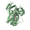
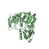
| Structure viewer | Molecule: MolmilJmol/JSmol |
|---|
- Downloads & links
-Download
| PDBx/mmCIF format | 7ncf.cif.gz | 161.6 KB | Display | PDBx/mmCIF format |
|---|---|---|---|---|
| PDB format | pdb7ncf.ent.gz | 126.9 KB | Display | PDB format |
| PDBx/mmJSON format | 7ncf.json.gz | Tree view | PDBx/mmJSON format | |
| Others |  Other downloads Other downloads |
-Validation report
| Arichive directory | https://data.pdbj.org/pub/pdb/validation_reports/nc/7ncfftp://data.pdbj.org/pub/pdb/validation_reports/nc/7ncf | HTTPS FTP |
|---|
-Related structure data
| Related structure data |  6p5sS S: Starting model for refinement |
|---|---|
| Similar structure data |


-Links
 PDBj
PDBj


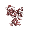
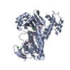
- Assembly
| Deposited unit | 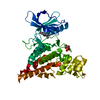
| ||||||||
|---|---|---|---|---|---|---|---|---|---|
| 1 |
| ||||||||
| Unit cell |
|
-Components
| #1: Protein | Mass: 42446.516 Da / Num. of mol.: 1 Source method: isolated from a genetically manipulated source Source: (gene. exp.) Homo sapiens (human) / Gene: HIPK2 / Plasmid: pNIC28-Bsa4 / Production host:  Escherichia coli BL21(DE3) (bacteria) / Variant (production host): -R3-pRARE2 Escherichia coli BL21(DE3) (bacteria) / Variant (production host): -R3-pRARE2References: UniProt: Q9H2X6, non-specific serine/threonine protein kinase |
|---|---|
| #2: Chemical | ChemComp-U82 /   Mass: 317.384 Da / Num. of mol.: 1 / Source method: obtained synthetically / Formula: C20H19N3O / Feature type: SUBJECT OF INVESTIGATION Mass: 317.384 Da / Num. of mol.: 1 / Source method: obtained synthetically / Formula: C20H19N3O / Feature type: SUBJECT OF INVESTIGATION |
| #3: Water | ChemComp-HOH / Water Mass: 18.015 Da / Num. of mol.: 23 / Source method: isolated from a natural source / Formula: H2O Mass: 18.015 Da / Num. of mol.: 23 / Source method: isolated from a natural source / Formula: H2O |
| Has ligand of interest | Y |
-Experimental details
-Experiment
| Experiment | Method: X-RAY DIFFRACTION / Number of used crystals: 1 |
|---|
- Sample preparation
| Crystal | Density Matthews: 3.46 Å3/Da / Density % sol: 64.44 % |
|---|---|
| Crystal grow | Temperature: 293.15 K / Method: vapor diffusion, sitting drop / pH: 7.5 Details: 10% v/v isopropanol, 0.2 M NaCl and 0.1 M HEPES, pH 7.5 |
-Data collection
| Diffraction | Mean temperature: 100 K / Serial crystal experiment: N | |||||||||||||||||||||||||||
|---|---|---|---|---|---|---|---|---|---|---|---|---|---|---|---|---|---|---|---|---|---|---|---|---|---|---|---|---|
| Diffraction source | Source: SYNCHROTRON / Site: SLS  / Beamline: X06SA / Wavelength: 1 Å / Beamline: X06SA / Wavelength: 1 Å | |||||||||||||||||||||||||||
| Detector | Type: DECTRIS EIGER2 X 16M / Detector: PIXEL / Date: Dec 18, 2020 | |||||||||||||||||||||||||||
| Radiation | Protocol: SINGLE WAVELENGTH / Monochromatic (M) / Laue (L): M / Scattering type: x-ray | |||||||||||||||||||||||||||
| Radiation wavelength | Wavelength: 1 Å / Relative weight: 1 | |||||||||||||||||||||||||||
| Reflection | Resolution: 2.72→43.81 Å / Num. obs: 15846 / % possible obs: 99.9 % / Redundancy: 8.8 % / CC1/2: 0.999 / Rmerge(I) obs: 0.073 / Rpim(I) all: 0.028 / Rrim(I) all: 0.082 / Net I/σ(I): 17.4 | |||||||||||||||||||||||||||
| Reflection shell | Diffraction-ID: 1
|
- Processing
| Software |
| |||||||||||||||||||||||||||||||||||||||||||||||||||||||||||||||||||||||||||
|---|---|---|---|---|---|---|---|---|---|---|---|---|---|---|---|---|---|---|---|---|---|---|---|---|---|---|---|---|---|---|---|---|---|---|---|---|---|---|---|---|---|---|---|---|---|---|---|---|---|---|---|---|---|---|---|---|---|---|---|---|---|---|---|---|---|---|---|---|---|---|---|---|---|---|---|---|
| Refinement | Method to determine structure: MOLECULAR REPLACEMENT Starting model: 6p5s Resolution: 2.72→43.81 Å / Cor.coef. Fo:Fc: 0.955 / Cor.coef. Fo:Fc free: 0.919 / SU B: 24.449 / SU ML: 0.231 / SU R Cruickshank DPI: 0.5011 / Cross valid method: THROUGHOUT / σ(F): 0 / ESU R: 0.501 / ESU R Free: 0.306 / Stereochemistry target values: MAXIMUM LIKELIHOOD Details: U VALUES : WITH TLS ADDED HYDROGENS HAVE BEEN ADDED IN THE RIDING POSITIONS
| |||||||||||||||||||||||||||||||||||||||||||||||||||||||||||||||||||||||||||
| Solvent computation | Ion probe radii: 0.8 Å / Shrinkage radii: 0.8 Å / VDW probe radii: 1.2 Å / Solvent model: MASK | |||||||||||||||||||||||||||||||||||||||||||||||||||||||||||||||||||||||||||
| Displacement parameters | Biso max: 192.85 Å2 / Biso mean: 89.316 Å2 / Biso min: 54.43 Å2
| |||||||||||||||||||||||||||||||||||||||||||||||||||||||||||||||||||||||||||
| Refinement step | Cycle: final / Resolution: 2.72→43.81 Å
| |||||||||||||||||||||||||||||||||||||||||||||||||||||||||||||||||||||||||||
| Refine LS restraints |
| |||||||||||||||||||||||||||||||||||||||||||||||||||||||||||||||||||||||||||
| LS refinement shell | Resolution: 2.72→2.79 Å / Rfactor Rfree error: 0 / Total num. of bins used: 20
| |||||||||||||||||||||||||||||||||||||||||||||||||||||||||||||||||||||||||||
| Refinement TLS params. | Method: refined / Refine-ID: X-RAY DIFFRACTION
| |||||||||||||||||||||||||||||||||||||||||||||||||||||||||||||||||||||||||||
| Refinement TLS group |
|
