 Movie
Movie Controller

[English] 日本語
 Yorodumi
- PDB-7nb3: Crystal structure of human choline alpha in complex with an inhibitor -
+ Open data

- Basic information
| Entry | Database: PDB / ID: 7nb3 | ||||||
|---|---|---|---|---|---|---|---|
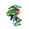
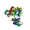
| Title | Crystal structure of human choline alpha in complex with an inhibitor | ||||||
 Components Components | Choline kinase alpha | ||||||
 Keywords Keywords |  TRANSFERASE / PHOSPHATIDYLCHOLINE / INHIBITOR COMPLEX TRANSFERASE / PHOSPHATIDYLCHOLINE / INHIBITOR COMPLEX | ||||||
| Function / homology |  Function and homology informationethanolamine kinase / choline kinase / ethanolamine kinase activity / CDP-choline pathway / choline kinase activity / Synthesis of PE / phosphatidylethanolamine biosynthetic process / lipid droplet disassembly / phosphatidylcholine biosynthetic process / cholinesterase activity ...ethanolamine kinase / choline kinase / ethanolamine kinase activity / CDP-choline pathway / choline kinase activity / Synthesis of PE / phosphatidylethanolamine biosynthetic process / lipid droplet disassembly / phosphatidylcholine biosynthetic process / cholinesterase activity / lipid transport / Synthesis of PC / cellular response to glucose starvation / lipid droplet / lipid metabolic process / protein tyrosine kinase activity / phosphorylation / protein homodimerization activity / ATP binding / cytosol / cytoplasm Function and homology informationethanolamine kinase / choline kinase / ethanolamine kinase activity / CDP-choline pathway / choline kinase activity / Synthesis of PE / phosphatidylethanolamine biosynthetic process / lipid droplet disassembly / phosphatidylcholine biosynthetic process / cholinesterase activity ...ethanolamine kinase / choline kinase / ethanolamine kinase activity / CDP-choline pathway / choline kinase activity / Synthesis of PE / phosphatidylethanolamine biosynthetic process / lipid droplet disassembly / phosphatidylcholine biosynthetic process / cholinesterase activity / lipid transport / Synthesis of PC / cellular response to glucose starvation / lipid droplet / lipid metabolic process / protein tyrosine kinase activity / phosphorylation / protein homodimerization activity / ATP binding / cytosol / cytoplasmSimilarity search - Function | ||||||
| Biological species |  Homo sapiens (human) Homo sapiens (human) | ||||||
| Method | X-RAY DIFFRACTION / SYNCHROTRON / MOLECULAR REPLACEMENT / Resolution: 2 Å | ||||||
 Authors Authors | Casale, E. / Fasolini, M. | ||||||
 Citation Citation | Journal: Bioorg.Med.Chem.Lett. / Year: 2021 Title: Identification of unprecedented ATP-competitive choline kinase inhibitors. Authors: Quartieri, F. / Nesi, M. / Avanzi, N.R. / Borghi, D. / Casale, E. / Corti, E. / Cucchi, U. / Donati, D. / Fasolini, M. / Felder, E.R. / Galvani, A. / Giorgini, M.L. / Lomolino, A. / ...Authors: Quartieri, F. / Nesi, M. / Avanzi, N.R. / Borghi, D. / Casale, E. / Corti, E. / Cucchi, U. / Donati, D. / Fasolini, M. / Felder, E.R. / Galvani, A. / Giorgini, M.L. / Lomolino, A. / Menichincheri, M. / Orrenius, C. / Perrera, C. / Re Depaolini, S. / Riccardi-Sirtori, F. / Salsi, E. / Isacchi, A. / Gnocchi, P. | ||||||
| History |
|
- Structure visualization
| Structure viewer | Molecule: MolmilJmol/JSmol |
|---|
- Downloads & links
-Download
| PDBx/mmCIF format | 7nb3.cif.gz | 172.1 KB | Display | PDBx/mmCIF format |
|---|---|---|---|---|
| PDB format | pdb7nb3.ent.gz | Display | PDB format | |
| PDBx/mmJSON format | 7nb3.json.gz | Tree view | PDBx/mmJSON format | |
| Others |  Other downloads Other downloads |
-Validation report
| Arichive directory | https://data.pdbj.org/pub/pdb/validation_reports/nb/7nb3ftp://data.pdbj.org/pub/pdb/validation_reports/nb/7nb3 | HTTPS FTP |
|---|
-Related structure data
| Related structure data |  7nb1C 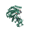 7nb2C  2ckqS S: Starting model for refinement C: citing same article ( |
|---|---|
| Similar structure data |


-Links
 PDBj
PDBj

- Assembly
| Deposited unit | 
| ||||||||
|---|---|---|---|---|---|---|---|---|---|
| 1 | 
| ||||||||
| 2 | 
| ||||||||
| Unit cell |
|
-Components
| #1: Protein | Mass: 45011.512 Da / Num. of mol.: 2 Source method: isolated from a genetically manipulated source Details: GLY 74 IS AN EXPRESSION TAG / Source: (gene. exp.) Homo sapiens (human) / Gene: CHKA, CHK, CKI / Production host:  Escherichia coli BL21(DE3) (bacteria) / Variant (production host): pLysS Escherichia coli BL21(DE3) (bacteria) / Variant (production host): pLysSReferences: UniProt: P35790, choline kinase, ethanolamine kinase#2: Chemical |   Mass: 443.501 Da / Num. of mol.: 2 / Source method: obtained synthetically / Formula: C24H25N7O2 / Feature type: SUBJECT OF INVESTIGATION Mass: 443.501 Da / Num. of mol.: 2 / Source method: obtained synthetically / Formula: C24H25N7O2 / Feature type: SUBJECT OF INVESTIGATION#3: Chemical |   Mass: 24.305 Da / Num. of mol.: 2 / Source method: obtained synthetically / Formula: Mg Mass: 24.305 Da / Num. of mol.: 2 / Source method: obtained synthetically / Formula: Mg#4: Chemical | HEPES  Mass: 238.305 Da / Num. of mol.: 3 / Source method: obtained synthetically / Formula: C8H18N2O4S / Comment: pH buffer*YM Mass: 238.305 Da / Num. of mol.: 3 / Source method: obtained synthetically / Formula: C8H18N2O4S / Comment: pH buffer*YM#5: Water | ChemComp-HOH / | Water Mass: 18.015 Da / Num. of mol.: 283 / Source method: isolated from a natural source / Formula: H2O Mass: 18.015 Da / Num. of mol.: 283 / Source method: isolated from a natural source / Formula: H2OHas ligand of interest | Y | |
|---|
-Experimental details
-Experiment
| Experiment | Method: X-RAY DIFFRACTION / Number of used crystals: 1 |
|---|
- Sample preparation
| Crystal | Density Matthews: 2.45 Å3/Da / Density % sol: 49.78 % |
|---|---|
| Crystal grow | Temperature: 293 K / Method: vapor diffusion, sitting drop / pH: 7 Details: 18-20% Peg-mme 5000, 0.1 M Magnesium Formate, 0.1 M HEPES pH 7.0 |
-Data collection
| Diffraction | Mean temperature: 100 K / Serial crystal experiment: N |
|---|---|
| Diffraction source | Source: SYNCHROTRON / Site: ESRF  / Beamline: ID23-1 / Wavelength: 0.976 Å / Beamline: ID23-1 / Wavelength: 0.976 Å |
| Detector | Type: DECTRIS PILATUS 6M-F / Detector: PIXEL / Date: Apr 23, 2015 |
| Radiation | Protocol: SINGLE WAVELENGTH / Monochromatic (M) / Laue (L): M / Scattering type: x-ray |
| Radiation wavelength | Wavelength: 0.976 Å / Relative weight: 1 |
| Reflection | Resolution: 2→47.3 Å / Num. obs: 60320 / % possible obs: 99.6 % / Redundancy: 4.4 % / Rmerge(I) obs: 0.054 / Net I/σ(I): 17.3 |
| Reflection shell | Resolution: 2→2.11 Å / Redundancy: 4.3 % / Rmerge(I) obs: 0.625 / Mean I/σ(I) obs: 2.3 / Num. unique obs: 8653 / % possible all: 99.6 |
- Processing
| Software |
| ||||||||||||||||||||||||||||||||||||||||||||||||||||||||||||||||||||||||||||||||||||||||||||||||||||||||||||||||||||||||||||||||||||||||||||
|---|---|---|---|---|---|---|---|---|---|---|---|---|---|---|---|---|---|---|---|---|---|---|---|---|---|---|---|---|---|---|---|---|---|---|---|---|---|---|---|---|---|---|---|---|---|---|---|---|---|---|---|---|---|---|---|---|---|---|---|---|---|---|---|---|---|---|---|---|---|---|---|---|---|---|---|---|---|---|---|---|---|---|---|---|---|---|---|---|---|---|---|---|---|---|---|---|---|---|---|---|---|---|---|---|---|---|---|---|---|---|---|---|---|---|---|---|---|---|---|---|---|---|---|---|---|---|---|---|---|---|---|---|---|---|---|---|---|---|---|---|---|
| Refinement | Method to determine structure: MOLECULAR REPLACEMENT Starting model: 2CKQ Resolution: 2→41.128 Å / Cor.coef. Fo:Fc: 0.961 / Cor.coef. Fo:Fc free: 0.939 / WRfactor Rfree: 0.222 / WRfactor Rwork: 0.175 / SU B: 4.612 / SU ML: 0.124 / Average fsc free: 0.8958 / Average fsc work: 0.9108 / Cross valid method: NONE / ESU R: 0.168 / ESU R Free: 0.157 Details: Hydrogens have been used if present in the input file
| ||||||||||||||||||||||||||||||||||||||||||||||||||||||||||||||||||||||||||||||||||||||||||||||||||||||||||||||||||||||||||||||||||||||||||||
| Solvent computation | Ion probe radii: 0.8 Å / Shrinkage radii: 0.8 Å / VDW probe radii: 1.2 Å / Solvent model: MASK BULK SOLVENT | ||||||||||||||||||||||||||||||||||||||||||||||||||||||||||||||||||||||||||||||||||||||||||||||||||||||||||||||||||||||||||||||||||||||||||||
| Displacement parameters | Biso mean: 40.519 Å2
| ||||||||||||||||||||||||||||||||||||||||||||||||||||||||||||||||||||||||||||||||||||||||||||||||||||||||||||||||||||||||||||||||||||||||||||
| Refinement step | Cycle: LAST / Resolution: 2→41.128 Å
| ||||||||||||||||||||||||||||||||||||||||||||||||||||||||||||||||||||||||||||||||||||||||||||||||||||||||||||||||||||||||||||||||||||||||||||
| Refine LS restraints |
| ||||||||||||||||||||||||||||||||||||||||||||||||||||||||||||||||||||||||||||||||||||||||||||||||||||||||||||||||||||||||||||||||||||||||||||
| LS refinement shell |
|
