 Movie
Movie Controller


+ Open data

- Basic information
| Entry | Database: PDB / ID: 7n7p | ||||||
|---|---|---|---|---|---|---|---|
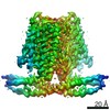
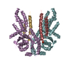
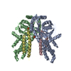
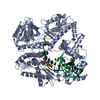
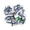
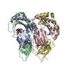
| Title | Cryo-EM structure of human TMEM120A | ||||||
 Components Components | Ion channel TACAN | ||||||
 Keywords Keywords |  MEMBRANE PROTEIN / Transmembrane protein MEMBRANE PROTEIN / Transmembrane protein | ||||||
| Function / homology |  Function and homology information Function and homology informationprotein heterooligomerization / nuclear inner membrane / fat cell differentiation / detection of mechanical stimulus involved in sensory perception of pain / monoatomic ion transmembrane transport / protein homooligomerization / monoatomic ion channel activity / membrane / plasma membraneSimilarity search - Function | ||||||
| Biological species |  Homo sapiens (human) Homo sapiens (human) | ||||||
| Method | ELECTRON MICROSCOPY / single particle reconstruction / cryo EM / Resolution: 3.24 Å | ||||||
 Authors Authors | Xue, J. / Han, Y. / Jiang, Y. | ||||||
| Funding support |  United States, 1items United States, 1items
| ||||||
 Citation Citation | Journal: Elife / Year: 2021 Title: TMEM120A is a coenzyme A-binding membrane protein with structural similarities to ELOVL fatty acid elongase. Authors: Jing Xue / Yan Han / Hamid Baniasadi / Weizhong Zeng / Jimin Pei / Nick V Grishin / Junmei Wang / Benjamin P Tu / Youxing Jiang / Abstract: TMEM120A, also named as TACAN, is a novel membrane protein highly conserved in vertebrates and was recently proposed to be a mechanosensitive channel involved in sensing mechanical pain. Here we ...TMEM120A, also named as TACAN, is a novel membrane protein highly conserved in vertebrates and was recently proposed to be a mechanosensitive channel involved in sensing mechanical pain. Here we present the single-particle cryogenic electron microscopy (cryo-EM) structure of human TMEM120A, which forms a tightly packed dimer with extensive interactions mediated by the N-terminal coiled coil domain (CCD), the C-terminal transmembrane domain (TMD), and the re-entrant loop between the two domains. The TMD of each TMEM120A subunit contains six transmembrane helices (TMs) and has no clear structural feature of a channel protein. Instead, the six TMs form an α-barrel with a deep pocket where a coenzyme A (CoA) molecule is bound. Intriguingly, some structural features of TMEM120A resemble those of elongase for very long-chain fatty acids (ELOVL) despite the low sequence homology between them, pointing to the possibility that TMEM120A may function as an enzyme for fatty acid metabolism, rather than a mechanosensitive channel. | ||||||
| History |
|
- Structure visualization
| Movie |
Movie viewer |
|---|---|
| Structure viewer | Molecule: MolmilJmol/JSmol |
- Downloads & links
-Download
| PDBx/mmCIF format | 7n7p.cif.gz | 121.5 KB | Display | PDBx/mmCIF format |
|---|---|---|---|---|
| PDB format | pdb7n7p.ent.gz | 100.4 KB | Display | PDB format |
| PDBx/mmJSON format | 7n7p.json.gz | Tree view | PDBx/mmJSON format | |
| Others |  Other downloads Other downloads |
-Validation report
| Arichive directory | https://data.pdbj.org/pub/pdb/validation_reports/n7/7n7pftp://data.pdbj.org/pub/pdb/validation_reports/n7/7n7p | HTTPS FTP |
|---|
-Related structure data
| Related structure data |  24230MC M: map data used to model this data C: citing same article ( |
|---|---|
| Similar structure data |


-Links
 PDBj
PDBj


- Assembly
| Deposited unit | 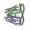
|
|---|---|
| 1 |
|
-Components
| #1: Protein | Mass: 42013.457 Da / Num. of mol.: 2 Source method: isolated from a genetically manipulated source Source: (gene. exp.) Homo sapiens (human) / Gene: TMEM120A, TACAN, TMPIT / Production host: Homo sapiens (human) / References: UniProt: Q9BXJ8#2: Chemical | Coenzyme A  Mass: 767.534 Da / Num. of mol.: 2 / Source method: obtained synthetically / Formula: C21H36N7O16P3S / Feature type: SUBJECT OF INVESTIGATION Mass: 767.534 Da / Num. of mol.: 2 / Source method: obtained synthetically / Formula: C21H36N7O16P3S / Feature type: SUBJECT OF INVESTIGATIONHas ligand of interest | Y | |
|---|
-Experimental details
-Experiment
| Experiment | Method: ELECTRON MICROSCOPY |
|---|---|
| EM experiment | Aggregation state: PARTICLE / 3D reconstruction method: single particle reconstruction |
- Sample preparation
| Component | Name: Human TMEM120A with CoA / Type: COMPLEX / Entity ID: #1 / Source: RECOMBINANT |
|---|---|
| Source (natural) | Organism: Homo sapiens (human) |
| Source (recombinant) | Organism: Homo sapiens (human) |
| Buffer solution | pH: 7.4 |
| Specimen | Embedding applied: NO / Shadowing applied: NO / Staining applied: NO / Vitrification applied: YES |
| Vitrification | Cryogen name: ETHANE |
- Electron microscopy imaging
| Experimental equipment |  Model: Titan Krios / Image courtesy: FEI Company |
|---|---|
| Microscopy | Model: FEI TITAN KRIOS |
| Electron gun | Electron source: FIELD EMISSION GUN / Accelerating voltage: 300 kV / Illumination mode: FLOOD BEAM |
| Electron lens | Mode: BRIGHT FIELDBright-field microscopy |
| Image recording | Electron dose: 1 e/Å2 / Film or detector model: GATAN K3 (6k x 4k) |
- Processing
| CTF correction | Type: PHASE FLIPPING ONLY |
|---|---|
| 3D reconstruction | Resolution: 3.24 Å / Resolution method: FSC 0.143 CUT-OFF / Num. of particles: 701283 / Symmetry type: POINT |
