 Movie
Movie Controller

[English] 日本語
 Yorodumi
- PDB-7n0w: Rigidity of loop 1 contributes to equipotency of globular and rib... -
+ Open data

- Basic information
| Entry | Database: PDB / ID: 7n0w | ||||||
|---|---|---|---|---|---|---|---|
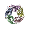
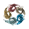
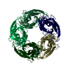
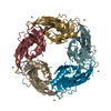
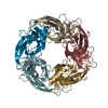
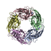
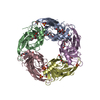
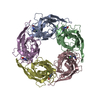
| Title | Rigidity of loop 1 contributes to equipotency of globular and ribbon isomers of alpha-conotoxin AusIA | ||||||
 Components Components |
| ||||||
 Keywords Keywords | CHOLINE-BINDING PROTEIN/ANTAGONIST / Alpha-conotoxin /  Complex / Acetylcholine-binding protein / CHOLINE-BINDING PROTEIN / CHOLINE-BINDING PROTEIN-ANTAGONIST complex Complex / Acetylcholine-binding protein / CHOLINE-BINDING PROTEIN / CHOLINE-BINDING PROTEIN-ANTAGONIST complex | ||||||
| Function / homology |  Function and homology informationacetylcholine receptor activity / acetylcholine-gated monoatomic cation-selective channel activity / synaptic cleft / response to nicotine / neuron projection / synapse / membrane Function and homology informationacetylcholine receptor activity / acetylcholine-gated monoatomic cation-selective channel activity / synaptic cleft / response to nicotine / neuron projection / synapse / membraneSimilarity search - Function | ||||||
| Biological species |   Lymnaea stagnalis (great pond snail)Conus australis (invertebrata) Lymnaea stagnalis (great pond snail)Conus australis (invertebrata) | ||||||
| Method | X-RAY DIFFRACTION / SYNCHROTRON / MOLECULAR REPLACEMENT / Resolution: 2.46 Å | ||||||
 Authors Authors | Ho, T.N.T. / Abraham, N. / Lewis, R.J. | ||||||
| Funding support |  Australia, 1items Australia, 1items
| ||||||
 Citation Citation | Journal: Sci Rep / Year: 2021 Title: Rigidity of loop 1 contributes to equipotency of globular and ribbon isomers of alpha-conotoxin AusIA. Authors: Ho, T.N.T. / Abraham, N. / Lewis, R.J. | ||||||
| History |
|
- Structure visualization
| Structure viewer | Molecule: MolmilJmol/JSmol |
|---|
- Downloads & links
-Download
| PDBx/mmCIF format | 7n0w.cif.gz | 420.3 KB | Display | PDBx/mmCIF format |
|---|---|---|---|---|
| PDB format | pdb7n0w.ent.gz | 351.2 KB | Display | PDB format |
| PDBx/mmJSON format | 7n0w.json.gz | Tree view | PDBx/mmJSON format | |
| Others |  Other downloads Other downloads |
-Validation report
| Arichive directory | https://data.pdbj.org/pub/pdb/validation_reports/n0/7n0wftp://data.pdbj.org/pub/pdb/validation_reports/n0/7n0w | HTTPS FTP |
|---|
-Related structure data
| Related structure data |  7n0yC  5t90S S: Starting model for refinement C: citing same article ( |
|---|---|
| Similar structure data |


-Links
 PDBj
PDBj

- Assembly
| Deposited unit | 
| ||||||||
|---|---|---|---|---|---|---|---|---|---|
| 1 |
| ||||||||
| Unit cell |
|
-Components
| #1: Protein | Mass: 23262.818 Da / Num. of mol.: 5 Source method: isolated from a genetically manipulated source Source: (gene. exp.) Lymnaea stagnalis (great pond snail) / Production host:  Escherichia coli (E. coli) / References: UniProt: P58154 Escherichia coli (E. coli) / References: UniProt: P58154#2: Protein/peptide | | Mass: 1772.067 Da / Num. of mol.: 1 / Source method: obtained synthetically / Source: (synth.) Conus australis (invertebrata)#3: Water | ChemComp-HOH / | Water Mass: 18.015 Da / Num. of mol.: 67 / Source method: isolated from a natural source / Formula: H2O Mass: 18.015 Da / Num. of mol.: 67 / Source method: isolated from a natural source / Formula: H2O |
|---|
-Experimental details
-Experiment
| Experiment | Method: X-RAY DIFFRACTION / Number of used crystals: 1 |
|---|
- Sample preparation
| Crystal | Density Matthews: 2.54 Å3/Da / Density % sol: 51.56 % |
|---|---|
| Crystal grow | Temperature: 296.15 K / Method: vapor diffusion, hanging drop Details: 0.1 M calcium acetate hydrate, 12% PEG400, 0.1M MES pH 6.0 |
-Data collection
| Diffraction | Mean temperature: 100 K / Serial crystal experiment: N |
|---|---|
| Diffraction source | Source: SYNCHROTRON / Site: Australian Synchrotron / Beamline: MX2 / Wavelength: 0.95373 Å |
| Detector | Type: DECTRIS EIGER X 16M / Detector: PIXEL / Date: Dec 3, 2019 |
| Radiation | Protocol: SINGLE WAVELENGTH / Monochromatic (M) / Laue (L): M / Scattering type: x-ray |
| Radiation wavelength | Wavelength: 0.95373 Å / Relative weight: 1 |
| Reflection | Resolution: 2.459→48.3 Å / Num. obs: 44280 / % possible obs: 99.2 % / Redundancy: 20 % / Rsym value: 0.076 / Net I/σ(I): 19.5 |
| Reflection shell | Resolution: 2.46→2.55 Å / Mean I/σ(I) obs: 2.2 / Num. unique obs: 4459 / Rsym value: 1.08 |
- Processing
| Software |
| |||||||||||||||||||||||||||||||||||||||||||||||||||||||||||||||||||||||||||||||||||||||||||||||||||||||||||||||||||||||||||||||||||||||||||||||||||||||||||||||||||||||||||||||
|---|---|---|---|---|---|---|---|---|---|---|---|---|---|---|---|---|---|---|---|---|---|---|---|---|---|---|---|---|---|---|---|---|---|---|---|---|---|---|---|---|---|---|---|---|---|---|---|---|---|---|---|---|---|---|---|---|---|---|---|---|---|---|---|---|---|---|---|---|---|---|---|---|---|---|---|---|---|---|---|---|---|---|---|---|---|---|---|---|---|---|---|---|---|---|---|---|---|---|---|---|---|---|---|---|---|---|---|---|---|---|---|---|---|---|---|---|---|---|---|---|---|---|---|---|---|---|---|---|---|---|---|---|---|---|---|---|---|---|---|---|---|---|---|---|---|---|---|---|---|---|---|---|---|---|---|---|---|---|---|---|---|---|---|---|---|---|---|---|---|---|---|---|---|---|---|---|
| Refinement | Method to determine structure: MOLECULAR REPLACEMENT Starting model: 5t90 Resolution: 2.46→48.3 Å / Cor.coef. Fo:Fc: 0.948 / Cor.coef. Fo:Fc free: 0.938 / SU B: 21.205 / SU ML: 0.218 / Cross valid method: THROUGHOUT / ESU R: 0.504 / ESU R Free: 0.267 / Stereochemistry target values: MAXIMUM LIKELIHOOD Details: U VALUES : WITH TLS ADDED HYDROGENS HAVE BEEN ADDED IN THE RIDING POSITIONS U VALUES : RESIDUAL ONLY
| |||||||||||||||||||||||||||||||||||||||||||||||||||||||||||||||||||||||||||||||||||||||||||||||||||||||||||||||||||||||||||||||||||||||||||||||||||||||||||||||||||||||||||||||
| Solvent computation | Ion probe radii: 0.9 Å / Shrinkage radii: 0.9 Å / VDW probe radii: 1.2 Å / Solvent model: MASK | |||||||||||||||||||||||||||||||||||||||||||||||||||||||||||||||||||||||||||||||||||||||||||||||||||||||||||||||||||||||||||||||||||||||||||||||||||||||||||||||||||||||||||||||
| Displacement parameters | Biso mean: 75.417 Å2
| |||||||||||||||||||||||||||||||||||||||||||||||||||||||||||||||||||||||||||||||||||||||||||||||||||||||||||||||||||||||||||||||||||||||||||||||||||||||||||||||||||||||||||||||
| Refinement step | Cycle: LAST / Resolution: 2.46→48.3 Å
| |||||||||||||||||||||||||||||||||||||||||||||||||||||||||||||||||||||||||||||||||||||||||||||||||||||||||||||||||||||||||||||||||||||||||||||||||||||||||||||||||||||||||||||||
| Refine LS restraints |
| |||||||||||||||||||||||||||||||||||||||||||||||||||||||||||||||||||||||||||||||||||||||||||||||||||||||||||||||||||||||||||||||||||||||||||||||||||||||||||||||||||||||||||||||
| LS refinement shell | Resolution: 2.46→2.523 Å
| |||||||||||||||||||||||||||||||||||||||||||||||||||||||||||||||||||||||||||||||||||||||||||||||||||||||||||||||||||||||||||||||||||||||||||||||||||||||||||||||||||||||||||||||
| Refinement TLS params. | Method: refined / Refine-ID: X-RAY DIFFRACTION
| |||||||||||||||||||||||||||||||||||||||||||||||||||||||||||||||||||||||||||||||||||||||||||||||||||||||||||||||||||||||||||||||||||||||||||||||||||||||||||||||||||||||||||||||
| Refinement TLS group |
|
