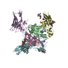
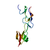
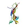
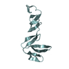
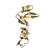
negative regulation of adaptive immune memory response / Costimulation by the CD28 family / negative regulation of alpha-beta T cell proliferation / TNFs bind their physiological receptors / positive regulation of cytokine production involved in immune response / cytokine binding / positive regulation of T cell migration / T cell costimulation / positive regulation of peptidyl-tyrosine phosphorylation / virus receptor activity ...negative regulation of adaptive immune memory response / Costimulation by the CD28 family / negative regulation of alpha-beta T cell proliferation / TNFs bind their physiological receptors / positive regulation of cytokine production involved in immune response / cytokine binding / positive regulation of T cell migration / T cell costimulation / positive regulation of peptidyl-tyrosine phosphorylation / virus receptor activity / adaptive immune response / defense response to Gram-negative bacterium / membrane => GO:0016020 / defense response to Gram-positive bacterium / external side of plasma membrane / innate immune response / ubiquitin protein ligase binding / plasma membrane Similarity search - Function
Tumour necrosis factor receptor 14 / Tumor necrosis factor receptor 14/UL144, N-terminal / TNFR/NGFR family cysteine-rich region domain profile. / TNFR/NGFR cysteine-rich region / TNFR/NGFR family cysteine-rich region signature. / Tumor necrosis factor receptor / nerve growth factor receptor repeats. / TNFR/NGFR cysteine-rich region Similarity search - Domain/homology
Resolution: 2.1→20 Å / Cor.coef. Fo:Fc: 0.956 / Cor.coef. Fo:Fc free: 0.933 / SU B: 6.702 / SU ML: 0.16 / SU R Cruickshank DPI: 0.182 / Cross valid method: THROUGHOUT / σ(F): 0 / ESU R: 0.182 / ESU R Free: 0.173 / Stereochemistry target values: MAXIMUM LIKELIHOOD Details: HYDROGENS HAVE BEEN ADDED IN THE RIDING POSITIONS U VALUES : REFINED INDIVIDUALLY
Rfactor
Num. reflection
% reflection
Selection details
Rfree
0.2575
464
5.2 %
RANDOM
Rwork
0.2119
-
-
-
obs
0.2142
8482
99.46 %
-
Solvent computation
Ion probe radii: 0.8 Å / Shrinkage radii: 0.8 Å / VDW probe radii: 1.2 Å / Solvent model: MASK
In the structure databanks used in Yorodumi, some data are registered as the other names, "COVID-19 virus" and "2019-nCoV". Here are the details of the virus and the list of structure data.
Jan 31, 2019. EMDB accession codes are about to change! (news from PDBe EMDB page)
EMDB accession codes are about to change! (news from PDBe EMDB page)
The allocation of 4 digits for EMDB accession codes will soon come to an end. Whilst these codes will remain in use, new EMDB accession codes will include an additional digit and will expand incrementally as the available range of codes is exhausted. The current 4-digit format prefixed with “EMD-” (i.e. EMD-XXXX) will advance to a 5-digit format (i.e. EMD-XXXXX), and so on. It is currently estimated that the 4-digit codes will be depleted around Spring 2019, at which point the 5-digit format will come into force.
The EM Navigator/Yorodumi systems omit the EMD- prefix.
Related info.:Q: What is EMD? / ID/Accession-code notation in Yorodumi/EM Navigator
Yorodumi is a browser for structure data from EMDB, PDB, SASBDB, etc.
This page is also the successor to EM Navigator detail page, and also detail information page/front-end page for Omokage search.
The word "yorodu" (or yorozu) is an old Japanese word meaning "ten thousand". "mi" (miru) is to see.
Related info.:EMDB / PDB / SASBDB / Comparison of 3 databanks / Yorodumi Search / Aug 31, 2016. New EM Navigator & Yorodumi / Yorodumi Papers / Jmol/JSmol / Function and homology information / Changes in new EM Navigator and Yorodumi
 Movie
Movie Controller


 Open data
 Basic information
 Components
 Keywords
 IMMUNE SYSTEM /
  Function and homology information
 Authors
 United States, 11items
  Citation
 Structure visualization
 Downloads & links
 Other downloads
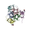


 PDBj
PDBj
 Assembly


 Mass: 96.063 Da / Num. of mol.: 3 / Source method: obtained synthetically / Formula: SO4
 Mass: 18.015 Da / Num. of mol.: 31 / Source method: isolated from a natural source / Formula: H2O
 Sample preparation
 Processing