 Movie
Movie Controller


+ Open data

- Basic information
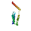
| Entry | Database: PDB / ID: 4fhq | ||||||
|---|---|---|---|---|---|---|---|
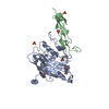
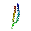
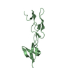
| Title | Crystal Structure of HVEM | ||||||
 Components Components | Tumor necrosis factor receptor superfamily member 14 | ||||||
 Keywords Keywords |  IMMUNE SYSTEM / CYSTEINE RICH DOMAIN / TNF RECEPTOR / Structural Genomics / PSI-Biology / Protein Structure Initiative / Atoms-to-Animals: The Immune Function Network / IFN / TNFRSF / Cysteine Rich Domains / Receptor / TNF14 / BTLA / CD160 / gD of HSV / Membrane / New York Structural Genomics Research Consortium (NYSGRC) IMMUNE SYSTEM / CYSTEINE RICH DOMAIN / TNF RECEPTOR / Structural Genomics / PSI-Biology / Protein Structure Initiative / Atoms-to-Animals: The Immune Function Network / IFN / TNFRSF / Cysteine Rich Domains / Receptor / TNF14 / BTLA / CD160 / gD of HSV / Membrane / New York Structural Genomics Research Consortium (NYSGRC) | ||||||
| Function / homology |  Function and homology information Function and homology informationnegative regulation of adaptive immune memory response / negative regulation of alpha-beta T cell proliferation / tumor necrosis factor receptor activity / positive regulation of cytokine production involved in immune response / TNFs bind their physiological receptors / Costimulation by the CD28 family / cytokine binding / positive regulation of T cell migration / T cell costimulation / positive regulation of peptidyl-tyrosine phosphorylation ...negative regulation of adaptive immune memory response / negative regulation of alpha-beta T cell proliferation / tumor necrosis factor receptor activity / positive regulation of cytokine production involved in immune response / TNFs bind their physiological receptors / Costimulation by the CD28 family / cytokine binding / positive regulation of T cell migration / T cell costimulation / positive regulation of peptidyl-tyrosine phosphorylation / virus receptor activity / adaptive immune response / defense response to Gram-negative bacterium / cell surface receptor signaling pathway / defense response to Gram-positive bacterium / immune response / external side of plasma membrane / innate immune response / ubiquitin protein ligase binding / plasma membraneSimilarity search - Function | ||||||
| Biological species |  Homo sapiens (human) Homo sapiens (human) | ||||||
| Method | X-RAY DIFFRACTION / SYNCHROTRON / MOLECULAR REPLACEMENT / Resolution: 2.251 Å | ||||||
 Authors Authors | Liu, W. / Zhan, C. / Patskovsky, Y. / Bhosle, R.C. / Nathenson, S.G. / Almo, S.C. / Atoms-to-Animals: The Immune Function Network (IFN) / New York Structural Genomics Research Consortium (NYSGRC) | ||||||
 Citation Citation | Journal: Mol Biotechnol / Year: 2015 Title: Increased Heterologous Protein Expression in Drosophila S2 Cells for Massive Production of Immune Ligands/Receptors and Structural Analysis of Human HVEM. Authors: Liu, W. / Vigdorovich, V. / Zhan, C. / Patskovsky, Y. / Bonanno, J.B. / Nathenson, S.G. / Almo, S.C. | ||||||
| History |
|
- Structure visualization
| Structure viewer | Molecule: MolmilJmol/JSmol |
|---|
- Downloads & links
-Download
| PDBx/mmCIF format | 4fhq.cif.gz | 51.9 KB | Display | PDBx/mmCIF format |
|---|---|---|---|---|
| PDB format | pdb4fhq.ent.gz | 37.2 KB | Display | PDB format |
| PDBx/mmJSON format | 4fhq.json.gz | Tree view | PDBx/mmJSON format | |
| Others |  Other downloads Other downloads |
-Validation report
| Arichive directory | https://data.pdbj.org/pub/pdb/validation_reports/fh/4fhqftp://data.pdbj.org/pub/pdb/validation_reports/fh/4fhq | HTTPS FTP |
|---|
-Related structure data
| Related structure data | 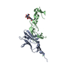 2aw2S S: Starting model for refinement |
|---|---|
| Similar structure data | |
| Other databases |


-Links
 PDBj
PDBj
- Assembly
| Deposited unit | 
| ||||||||
|---|---|---|---|---|---|---|---|---|---|
| 1 |
| ||||||||
| Unit cell |
| ||||||||
| Details | AUTHOR DETERMINED BIOLOGICAL UNIT: NOT DETERMINED |
-Components
| #1: Protein | Mass: 14332.176 Da / Num. of mol.: 1 / Fragment: extracellular domain residues 39-162 Source method: isolated from a genetically manipulated source Source: (gene. exp.) Homo sapiens (human) / Gene: HVEA, HVEM, TNFRSF14, UNQ329/PRO509 / Plasmid: pMT-Bip-His / Cell (production host): Schneider 2 / Production host:  Drosophila melanogaster (fruit fly) / References: UniProt: Q92956 Drosophila melanogaster (fruit fly) / References: UniProt: Q92956 |
|---|---|
| #2: Water | ChemComp-HOH / Water Mass: 18.015 Da / Num. of mol.: 2 / Source method: isolated from a natural source / Formula: H2O Mass: 18.015 Da / Num. of mol.: 2 / Source method: isolated from a natural source / Formula: H2O |
-Experimental details
-Experiment
| Experiment | Method: X-RAY DIFFRACTION / Number of used crystals: 1 |
|---|
- Sample preparation
| Crystal | Density Matthews: 3 Å3/Da / Density % sol: 58.99 % |
|---|---|
| Crystal grow | Temperature: 294 K / Method: vapor diffusion, sitting drop / pH: 8.5 Details: 0.2 M SODIUM ACETATE TRIHYDRATE, PH 8.5, TRIS-HCL, 30% W/V PEG4000, VAPOR DIFFUSION, SITTING DROP, temperature 294.0K |
-Data collection
| Diffraction | Mean temperature: 100 K |
|---|---|
| Diffraction source | Source: SYNCHROTRON / Site: NSLS  / Beamline: X29A / Wavelength: 1.075 / Wavelength: 1.075 Å / Beamline: X29A / Wavelength: 1.075 / Wavelength: 1.075 Å |
| Detector | Type: ADSC QUANTUM 315 / Detector: CCD / Date: Mar 15, 2012 / Details: MIRRORS |
| Radiation | Protocol: SINGLE WAVELENGTH / Monochromatic (M) / Laue (L): M / Scattering type: x-ray |
| Radiation wavelength | Wavelength: 1.075 Å / Relative weight: 1 |
| Reflection | Resolution: 2.25→50 Å / Num. obs: 8551 / % possible obs: 99.3 % / Redundancy: 7.6 % / Biso Wilson estimate: 54.17 Å2 / Rmerge(I) obs: 0.104 / Rsym value: 0.09 / Net I/σ(I): 16.68 |
| Reflection shell | Resolution: 2.25→2.29 Å / Redundancy: 6.4 % / Mean I/σ(I) obs: 1.062 / % possible all: 94.7 |
- Processing
| Software |
| |||||||||||||||||||||||||||||||||||||||||||||||||||||||||||||||||||||||||||
|---|---|---|---|---|---|---|---|---|---|---|---|---|---|---|---|---|---|---|---|---|---|---|---|---|---|---|---|---|---|---|---|---|---|---|---|---|---|---|---|---|---|---|---|---|---|---|---|---|---|---|---|---|---|---|---|---|---|---|---|---|---|---|---|---|---|---|---|---|---|---|---|---|---|---|---|---|
| Refinement | Method to determine structure: MOLECULAR REPLACEMENT Starting model: 2AW2 Resolution: 2.251→33.494 Å / SU ML: 0.47 / σ(F): 0 / Phase error: 38.13 / Stereochemistry target values: MLHL
| |||||||||||||||||||||||||||||||||||||||||||||||||||||||||||||||||||||||||||
| Solvent computation | Shrinkage radii: 0.6 Å / VDW probe radii: 0.9 Å / Solvent model: FLAT BULK SOLVENT MODEL / Bsol: 66.537 Å2 / ksol: 0.359 e/Å3 | |||||||||||||||||||||||||||||||||||||||||||||||||||||||||||||||||||||||||||
| Displacement parameters |
| |||||||||||||||||||||||||||||||||||||||||||||||||||||||||||||||||||||||||||
| Refinement step | Cycle: LAST / Resolution: 2.251→33.494 Å
| |||||||||||||||||||||||||||||||||||||||||||||||||||||||||||||||||||||||||||
| Refine LS restraints |
| |||||||||||||||||||||||||||||||||||||||||||||||||||||||||||||||||||||||||||
| LS refinement shell |
| |||||||||||||||||||||||||||||||||||||||||||||||||||||||||||||||||||||||||||
| Refinement TLS params. | Method: refined / Refine-ID: X-RAY DIFFRACTION
| |||||||||||||||||||||||||||||||||||||||||||||||||||||||||||||||||||||||||||
| Refinement TLS group |
|
