 Movie
Movie Controller


+ Open data

- Basic information
| Entry | Database: PDB / ID: 7mru | ||||||
|---|---|---|---|---|---|---|---|
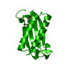
| Title | Crystal structure of S62A MIF2 mutant | ||||||
 Components Components | D-dopachrome decarboxylase | ||||||
 Keywords Keywords | CYTOKINE | ||||||
| Function / homology |  Function and homology informationD-dopachrome decarboxylase / D-dopachrome decarboxylase activity / melanin biosynthetic process / dopachrome isomerase activity / phenylpyruvate tautomerase activity / cytokine receptor binding / negative regulation of macrophage chemotaxis / positive regulation of inflammatory response / positive regulation of tumor necrosis factor production / positive regulation of ERK1 and ERK2 cascade ...D-dopachrome decarboxylase / D-dopachrome decarboxylase activity / melanin biosynthetic process / dopachrome isomerase activity / phenylpyruvate tautomerase activity / cytokine receptor binding / negative regulation of macrophage chemotaxis / positive regulation of inflammatory response / positive regulation of tumor necrosis factor production / positive regulation of ERK1 and ERK2 cascade / extracellular space / extracellular exosome / cytoplasm Function and homology informationD-dopachrome decarboxylase / D-dopachrome decarboxylase activity / melanin biosynthetic process / dopachrome isomerase activity / phenylpyruvate tautomerase activity / cytokine receptor binding / negative regulation of macrophage chemotaxis / positive regulation of inflammatory response / positive regulation of tumor necrosis factor production / positive regulation of ERK1 and ERK2 cascade ...D-dopachrome decarboxylase / D-dopachrome decarboxylase activity / melanin biosynthetic process / dopachrome isomerase activity / phenylpyruvate tautomerase activity / cytokine receptor binding / negative regulation of macrophage chemotaxis / positive regulation of inflammatory response / positive regulation of tumor necrosis factor production / positive regulation of ERK1 and ERK2 cascade / extracellular space / extracellular exosome / cytoplasmSimilarity search - Function | ||||||
| Biological species |  Homo sapiens (human) Homo sapiens (human) | ||||||
| Method | X-RAY DIFFRACTION / MOLECULAR REPLACEMENT / Resolution: 1.33 Å | ||||||
 Authors Authors | Murphy, E.L. / Manjula, R. / Murphy, J.W. / Lolis, E. | ||||||
 Citation Citation | Journal: J.Biol.Chem. / Year: 2021 Title: A structurally preserved allosteric site in the MIF superfamily affects enzymatic activity and CD74 activation in D-dopachrome tautomerase. Authors: Chen, E. / Reiss, K. / Shah, D. / Manjula, R. / Allen, B. / Murphy, E.L. / Murphy, J.W. / Batista, V.S. / Bhandari, V. / Lolis, E.J. / Lisi, G.P. | ||||||
| History |
|
- Structure visualization
| Structure viewer | Molecule: MolmilJmol/JSmol |
|---|
- Downloads & links
-Download
| PDBx/mmCIF format | 7mru.cif.gz | 96.1 KB | Display | PDBx/mmCIF format |
|---|---|---|---|---|
| PDB format | pdb7mru.ent.gz | 70.1 KB | Display | PDB format |
| PDBx/mmJSON format | 7mru.json.gz | Tree view | PDBx/mmJSON format | |
| Others |  Other downloads Other downloads |
-Validation report
| Arichive directory | https://data.pdbj.org/pub/pdb/validation_reports/mr/7mruftp://data.pdbj.org/pub/pdb/validation_reports/mr/7mru | HTTPS FTP |
|---|
-Related structure data
| Related structure data |  7mrvC  7mseC  7mw7C  3kanS S: Starting model for refinement C: citing same article ( |
|---|---|
| Similar structure data |


-Links
 PDBj
PDBj- Assembly
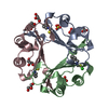
| Deposited unit | 
| ||||||||
|---|---|---|---|---|---|---|---|---|---|
| 1 |
| ||||||||
| Unit cell |
|
-Components
| #1: Protein | / D-dopachrome tautomerase / Phenylpyruvate tautomerase II Mass: 12577.526 Da / Num. of mol.: 3 / Mutation: S62A Source method: isolated from a genetically manipulated source Source: (gene. exp.) Homo sapiens (human) / Gene: DDT / Production host:  Escherichia coli (E. coli) / References: UniProt: P30046, D-dopachrome decarboxylase Escherichia coli (E. coli) / References: UniProt: P30046, D-dopachrome decarboxylase#2: Chemical | ChemComp-TRS / | Tris  Mass: 122.143 Da / Num. of mol.: 1 / Source method: obtained synthetically / Formula: C4H12NO3 / Feature type: SUBJECT OF INVESTIGATION / Comment: pH buffer*YM Mass: 122.143 Da / Num. of mol.: 1 / Source method: obtained synthetically / Formula: C4H12NO3 / Feature type: SUBJECT OF INVESTIGATION / Comment: pH buffer*YM#3: Water | ChemComp-HOH / | Water Mass: 18.015 Da / Num. of mol.: 623 / Source method: isolated from a natural source / Formula: H2O Mass: 18.015 Da / Num. of mol.: 623 / Source method: isolated from a natural source / Formula: H2OHas ligand of interest | Y | |
|---|
-Experimental details
-Experiment
| Experiment | Method: X-RAY DIFFRACTION / Number of used crystals: 1 |
|---|
- Sample preparation
| Crystal | Density Matthews: 2.13 Å3/Da / Density % sol: 42.2 % |
|---|---|
| Crystal grow | Temperature: 293 K / Method: vapor diffusion, hanging drop / pH: 8.5 / Details: 0.2 N NaCl, 25%w/v PEG 3350, 0.1 M Tris at pH 8.5 |
-Data collection
| Diffraction | Mean temperature: 100 K / Serial crystal experiment: N | |||||||||||||||||||||||||||||||||||||||||||||||||||||||||||||||||||||||||||||||||||||||||||||||||||||||||||||||||||||||||||||||||||||||||||||||||||||||||||||||||||||||||||||||||||||||||||||
|---|---|---|---|---|---|---|---|---|---|---|---|---|---|---|---|---|---|---|---|---|---|---|---|---|---|---|---|---|---|---|---|---|---|---|---|---|---|---|---|---|---|---|---|---|---|---|---|---|---|---|---|---|---|---|---|---|---|---|---|---|---|---|---|---|---|---|---|---|---|---|---|---|---|---|---|---|---|---|---|---|---|---|---|---|---|---|---|---|---|---|---|---|---|---|---|---|---|---|---|---|---|---|---|---|---|---|---|---|---|---|---|---|---|---|---|---|---|---|---|---|---|---|---|---|---|---|---|---|---|---|---|---|---|---|---|---|---|---|---|---|---|---|---|---|---|---|---|---|---|---|---|---|---|---|---|---|---|---|---|---|---|---|---|---|---|---|---|---|---|---|---|---|---|---|---|---|---|---|---|---|---|---|---|---|---|---|---|---|---|---|
| Diffraction source | Source: ROTATING ANODE / Type: RIGAKU MICROMAX-007 / Wavelength: 1.51418 Å | |||||||||||||||||||||||||||||||||||||||||||||||||||||||||||||||||||||||||||||||||||||||||||||||||||||||||||||||||||||||||||||||||||||||||||||||||||||||||||||||||||||||||||||||||||||||||||||
| Detector | Type: DECTRIS PILATUS 200K / Detector: PIXEL / Date: Oct 21, 2020 | |||||||||||||||||||||||||||||||||||||||||||||||||||||||||||||||||||||||||||||||||||||||||||||||||||||||||||||||||||||||||||||||||||||||||||||||||||||||||||||||||||||||||||||||||||||||||||||
| Radiation | Protocol: SINGLE WAVELENGTH / Monochromatic (M) / Laue (L): M / Scattering type: x-ray | |||||||||||||||||||||||||||||||||||||||||||||||||||||||||||||||||||||||||||||||||||||||||||||||||||||||||||||||||||||||||||||||||||||||||||||||||||||||||||||||||||||||||||||||||||||||||||||
| Radiation wavelength | Wavelength: 1.51418 Å / Relative weight: 1 | |||||||||||||||||||||||||||||||||||||||||||||||||||||||||||||||||||||||||||||||||||||||||||||||||||||||||||||||||||||||||||||||||||||||||||||||||||||||||||||||||||||||||||||||||||||||||||||
| Reflection | Resolution: 1.33→50 Å / Num. obs: 71882 / % possible obs: 95.1 % / Redundancy: 6.5 % / Rmerge(I) obs: 0.058 / Rpim(I) all: 0.022 / Rrim(I) all: 0.062 / Χ2: 0.961 / Net I/σ(I): 11.2 / Num. measured all: 468497 | |||||||||||||||||||||||||||||||||||||||||||||||||||||||||||||||||||||||||||||||||||||||||||||||||||||||||||||||||||||||||||||||||||||||||||||||||||||||||||||||||||||||||||||||||||||||||||||
| Reflection shell | Diffraction-ID: 1
|
- Processing
| Software |
| ||||||||||||||||||||||||||||||||||||||||||||||||||||||||||||||||||||||||||||||||||||||||||||||||||||||||||||||||||||||||||||||||||||||||||||||||||||||||||||||||||||||||||||||||||||||
|---|---|---|---|---|---|---|---|---|---|---|---|---|---|---|---|---|---|---|---|---|---|---|---|---|---|---|---|---|---|---|---|---|---|---|---|---|---|---|---|---|---|---|---|---|---|---|---|---|---|---|---|---|---|---|---|---|---|---|---|---|---|---|---|---|---|---|---|---|---|---|---|---|---|---|---|---|---|---|---|---|---|---|---|---|---|---|---|---|---|---|---|---|---|---|---|---|---|---|---|---|---|---|---|---|---|---|---|---|---|---|---|---|---|---|---|---|---|---|---|---|---|---|---|---|---|---|---|---|---|---|---|---|---|---|---|---|---|---|---|---|---|---|---|---|---|---|---|---|---|---|---|---|---|---|---|---|---|---|---|---|---|---|---|---|---|---|---|---|---|---|---|---|---|---|---|---|---|---|---|---|---|---|---|
| Refinement | Method to determine structure: MOLECULAR REPLACEMENT Starting model: 3KAN Resolution: 1.33→42.86 Å / SU ML: 0.11 / Cross valid method: THROUGHOUT / σ(F): 1.33 / Phase error: 17.71 / Stereochemistry target values: ML
| ||||||||||||||||||||||||||||||||||||||||||||||||||||||||||||||||||||||||||||||||||||||||||||||||||||||||||||||||||||||||||||||||||||||||||||||||||||||||||||||||||||||||||||||||||||||
| Solvent computation | Shrinkage radii: 0.9 Å / VDW probe radii: 1.11 Å / Solvent model: FLAT BULK SOLVENT MODEL | ||||||||||||||||||||||||||||||||||||||||||||||||||||||||||||||||||||||||||||||||||||||||||||||||||||||||||||||||||||||||||||||||||||||||||||||||||||||||||||||||||||||||||||||||||||||
| Displacement parameters | Biso max: 42.13 Å2 / Biso mean: 17.2115 Å2 / Biso min: 9.79 Å2 | ||||||||||||||||||||||||||||||||||||||||||||||||||||||||||||||||||||||||||||||||||||||||||||||||||||||||||||||||||||||||||||||||||||||||||||||||||||||||||||||||||||||||||||||||||||||
| Refinement step | Cycle: final / Resolution: 1.33→42.86 Å
| ||||||||||||||||||||||||||||||||||||||||||||||||||||||||||||||||||||||||||||||||||||||||||||||||||||||||||||||||||||||||||||||||||||||||||||||||||||||||||||||||||||||||||||||||||||||
| LS refinement shell | Refine-ID: X-RAY DIFFRACTION / Rfactor Rfree error: 0 / Total num. of bins used: 25
|
