 Movie
Movie Controller

[English] 日本語
 Yorodumi
- PDB-7mhe: Thioesterase Domain of Human Fatty Acid Synthase (FASN-TE) bindin... -
+ Open data

- Basic information
| Entry | Database: PDB / ID: 7mhe | ||||||
|---|---|---|---|---|---|---|---|
| Title | Thioesterase Domain of Human Fatty Acid Synthase (FASN-TE) binding a competitive inhibitor SBP-7957 | ||||||
 Components Components | Fatty acid synthase | ||||||
 Keywords Keywords | HYDROLASE/Inhibitor / THIOESTERASE DOMAIN / FATTY ACID SYNTHASE / FASN-TE / HYDROLASE-Inhibitor complex | ||||||
| Function / homology |  Function and homology information Function and homology informationfatty-acid synthase system / (3R)-3-hydroxybutanoyl-[acyl-carrier-protein] hydratase activity / fatty acyl-[ACP] hydrolase activity / ether lipid biosynthetic process / (3R)-3-hydroxyoctanoyl-[acyl-carrier-protein] dehydratase activity / Vitamin B5 (pantothenate) metabolism / neutrophil differentiation / enoyl-[acyl-carrier-protein] reductase (NADPH, Re-specific) / (3R)-3-hydroxydecanoyl-[acyl-carrier-protein] dehydratase activity / glandular epithelial cell development ...fatty-acid synthase system / (3R)-3-hydroxybutanoyl-[acyl-carrier-protein] hydratase activity / fatty acyl-[ACP] hydrolase activity / ether lipid biosynthetic process / (3R)-3-hydroxyoctanoyl-[acyl-carrier-protein] dehydratase activity / Vitamin B5 (pantothenate) metabolism / neutrophil differentiation / enoyl-[acyl-carrier-protein] reductase (NADPH, Re-specific) / (3R)-3-hydroxydecanoyl-[acyl-carrier-protein] dehydratase activity / glandular epithelial cell development / : / glycogen granule / establishment of endothelial intestinal barrier / [acyl-carrier-protein] S-acetyltransferase / [acyl-carrier-protein] S-acetyltransferase activity / Fatty acyl-CoA biosynthesis / oleoyl-[acyl-carrier-protein] hydrolase / (3R)-3-hydroxypalmitoyl-[acyl-carrier-protein] dehydratase activity / modulation by host of viral process / (3R)-3-hydroxymyristoyl-[acyl-carrier-protein] dehydratase activity / ChREBP activates metabolic gene expression / [acyl-carrier-protein] S-malonyltransferase / [acyl-carrier-protein] S-malonyltransferase activity / 3-hydroxyacyl-[acyl-carrier-protein] dehydratase / beta-ketoacyl-[acyl-carrier-protein] synthase I / NR1H2 & NR1H3 regulate gene expression linked to lipogenesis / mammary gland development / 3-oxoacyl-[acyl-carrier-protein] reductase / 3-oxoacyl-[acyl-carrier-protein] reductase (NADPH) activity / fatty acid synthase activity / phosphopantetheine binding / monocyte differentiation / 3-oxoacyl-[acyl-carrier-protein] synthase activity / cellular response to interleukin-4 / Activation of gene expression by SREBF (SREBP) / fatty acid metabolic process / fatty acid biosynthetic process / osteoblast differentiation / melanosome / cadherin binding / inflammatory response / Golgi apparatus / RNA binding / extracellular exosome / membrane / plasma membrane / cytosol / cytoplasmSimilarity search - Function | ||||||
| Biological species |  Homo sapiens (human) Homo sapiens (human) | ||||||
| Method | X-RAY DIFFRACTION / SYNCHROTRON / MOLECULAR REPLACEMENT / Resolution: 2.8 Å | ||||||
 Authors Authors | Aleshin, A.E. / Lambert, L. / Liddington, R.C. / Cosford, N. | ||||||
 Citation Citation | Journal: To Be Published Title: Thioesterase Domain of Human Fatty Acid Synthase (FASN-TE) binding a competitive inhibitor SBP-7635 Authors: Aleshin, A.E. / Lambert, L. / Liddington, R.C. / Cosford, N. | ||||||
| History |
|
- Structure visualization
| Structure viewer | Molecule: MolmilJmol/JSmol |
|---|
- Downloads & links
-Download
| PDBx/mmCIF format | 7mhe.cif.gz | 72.9 KB | Display | PDBx/mmCIF format |
|---|---|---|---|---|
| PDB format | pdb7mhe.ent.gz | Display | PDB format | |
| PDBx/mmJSON format | 7mhe.json.gz | Tree view | PDBx/mmJSON format | |
| Others |  Other downloads Other downloads |
-Validation report
| Arichive directory | https://data.pdbj.org/pub/pdb/validation_reports/mh/7mheftp://data.pdbj.org/pub/pdb/validation_reports/mh/7mhe | HTTPS FTP |
|---|
-Related structure data
| Related structure data | 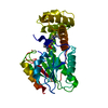 7mhdC 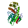 3tjmS C: citing same article ( S: Starting model for refinement |
|---|---|
| Similar structure data |
-Links
 PDBj
PDBj


- Assembly
| Deposited unit | 
| ||||||||
|---|---|---|---|---|---|---|---|---|---|
| 1 |
| ||||||||
| Unit cell |
|
-Components
| #1: Protein | / Type I fatty acid synthase Mass: 32524.670 Da / Num. of mol.: 1 / Fragment: Thioesterase Source method: isolated from a genetically manipulated source Source: (gene. exp.) Homo sapiens (human) / Gene: FASN, FAS / Production host:  Escherichia coli (E. coli) Escherichia coli (E. coli)References: UniProt: P49327, oleoyl-[acyl-carrier-protein] hydrolase |
|---|---|
| #2: Chemical | ChemComp-ZEG /   Mass: 404.478 Da / Num. of mol.: 1 / Source method: obtained synthetically / Formula: C19H17FN2O3S2 / Feature type: SUBJECT OF INVESTIGATION Mass: 404.478 Da / Num. of mol.: 1 / Source method: obtained synthetically / Formula: C19H17FN2O3S2 / Feature type: SUBJECT OF INVESTIGATION |
| #3: Chemical | ChemComp-EDO / Ethylene glycol  Mass: 62.068 Da / Num. of mol.: 1 / Source method: obtained synthetically / Formula: C2H6O2 Mass: 62.068 Da / Num. of mol.: 1 / Source method: obtained synthetically / Formula: C2H6O2 |
| #4: Water | ChemComp-HOH / Water Mass: 18.015 Da / Num. of mol.: 15 / Source method: isolated from a natural source / Formula: H2O Mass: 18.015 Da / Num. of mol.: 15 / Source method: isolated from a natural source / Formula: H2O |
| Has ligand of interest | Y |
-Experimental details
-Experiment
| Experiment | Method: X-RAY DIFFRACTION / Number of used crystals: 1 |
|---|
- Sample preparation
| Crystal | Density Matthews: 1.73 Å3/Da / Density % sol: 28.92 % |
|---|---|
| Crystal grow | Temperature: 293 K / Method: vapor diffusion, sitting drop Details: 0.3 uL of 8 mg/ml FAS-TE in 100 mM NaCl, 50 mM BisTris pH 6.0, 10 mM DTT, 0.5 mM of the inhibitor and 1% DMSO was mixed with 0.2 uL of well solution 10% PEG400, 50 mM Tris-Cl pH 8.5, 1.0 mM ...Details: 0.3 uL of 8 mg/ml FAS-TE in 100 mM NaCl, 50 mM BisTris pH 6.0, 10 mM DTT, 0.5 mM of the inhibitor and 1% DMSO was mixed with 0.2 uL of well solution 10% PEG400, 50 mM Tris-Cl pH 8.5, 1.0 mM DTT, 1.0 mM Ethylenediaminetetraacetic acid disodium salt (EDTA), 300 mM NaCl. PH range: 6.0-8.5 |
-Data collection
| Diffraction | Mean temperature: 100 K / Serial crystal experiment: N |
|---|---|
| Diffraction source | Source: SYNCHROTRON / Site: SSRL  / Beamline: BL9-2 / Wavelength: 1.03316 Å / Beamline: BL9-2 / Wavelength: 1.03316 Å |
| Detector | Type: DECTRIS PILATUS 6M / Detector: PIXEL / Date: Feb 9, 2016 |
| Radiation | Protocol: SINGLE WAVELENGTH / Monochromatic (M) / Laue (L): M / Scattering type: x-ray |
| Radiation wavelength | Wavelength: 1.03316 Å / Relative weight: 1 |
| Reflection | Resolution: 2.8→45.3 Å / Num. obs: 5295 / % possible obs: 90.7 % / Redundancy: 4.6 % / Rmerge(I) obs: 0.2 / Net I/σ(I): 5.2 |
| Reflection shell | Resolution: 2.8→2.9 Å / Rmerge(I) obs: 1.19 / Mean I/σ(I) obs: 1.4 / Num. unique obs: 759 |
- Processing
| Software |
| |||||||||||||||||||||||||||||||||||||||||||||||||||||||||||||||||||||||||||||||||||||||||||||||||||||||||||||||||||||||||||||||||||||||||||||||||||||||||||
|---|---|---|---|---|---|---|---|---|---|---|---|---|---|---|---|---|---|---|---|---|---|---|---|---|---|---|---|---|---|---|---|---|---|---|---|---|---|---|---|---|---|---|---|---|---|---|---|---|---|---|---|---|---|---|---|---|---|---|---|---|---|---|---|---|---|---|---|---|---|---|---|---|---|---|---|---|---|---|---|---|---|---|---|---|---|---|---|---|---|---|---|---|---|---|---|---|---|---|---|---|---|---|---|---|---|---|---|---|---|---|---|---|---|---|---|---|---|---|---|---|---|---|---|---|---|---|---|---|---|---|---|---|---|---|---|---|---|---|---|---|---|---|---|---|---|---|---|---|---|---|---|---|---|---|---|---|
| Refinement | Method to determine structure: MOLECULAR REPLACEMENT Starting model: 3TJM Resolution: 2.8→37.849 Å / Cor.coef. Fo:Fc: 0.939 / Cor.coef. Fo:Fc free: 0.876 / SU B: 25.392 / SU ML: 0.469 / Cross valid method: THROUGHOUT / ESU R Free: 0.558 Details: Hydrogens have been added in their riding positions
| |||||||||||||||||||||||||||||||||||||||||||||||||||||||||||||||||||||||||||||||||||||||||||||||||||||||||||||||||||||||||||||||||||||||||||||||||||||||||||
| Solvent computation | Ion probe radii: 0.8 Å / Shrinkage radii: 0.8 Å / VDW probe radii: 1.2 Å / Solvent model: MASK BULK SOLVENT | |||||||||||||||||||||||||||||||||||||||||||||||||||||||||||||||||||||||||||||||||||||||||||||||||||||||||||||||||||||||||||||||||||||||||||||||||||||||||||
| Displacement parameters | Biso mean: 59.85 Å2
| |||||||||||||||||||||||||||||||||||||||||||||||||||||||||||||||||||||||||||||||||||||||||||||||||||||||||||||||||||||||||||||||||||||||||||||||||||||||||||
| Refinement step | Cycle: LAST / Resolution: 2.8→37.849 Å
| |||||||||||||||||||||||||||||||||||||||||||||||||||||||||||||||||||||||||||||||||||||||||||||||||||||||||||||||||||||||||||||||||||||||||||||||||||||||||||
| Refine LS restraints |
| |||||||||||||||||||||||||||||||||||||||||||||||||||||||||||||||||||||||||||||||||||||||||||||||||||||||||||||||||||||||||||||||||||||||||||||||||||||||||||
| LS refinement shell | Resolution: 2.8→2.872 Å
|
