 Movie
Movie Controller

[English] 日本語
 Yorodumi
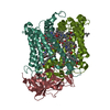
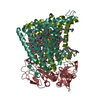
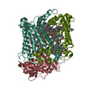
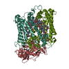
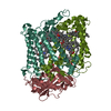
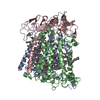
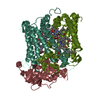
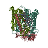
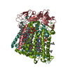
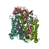
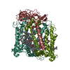
- PDB-7mh4: Crystal structure of R. sphaeroides Photosynthetic Reaction Cente... -
+ Open data

- Basic information
| Entry | Database: PDB / ID: 7mh4 | |||||||||
|---|---|---|---|---|---|---|---|---|---|---|
| Title | Crystal structure of R. sphaeroides Photosynthetic Reaction Center variant; Y(M210)3-bromotyrosine | |||||||||
 Components Components | (Reaction center protein ... Photosynthetic reaction centre) x 3 Photosynthetic reaction centre) x 3 | |||||||||
 Keywords Keywords | PHOTOSYNTHESIS / Photosynthetic / membrane protein / noncanonical amino acid / bromotyrosine | |||||||||
| Function / homology |  Function and homology information Function and homology informationplasma membrane-derived chromatophore membrane / plasma membrane light-harvesting complex / bacteriochlorophyll binding / photosynthesis, light reaction / electron transporter, transferring electrons within the cyclic electron transport pathway of photosynthesis activity / photosynthetic electron transport in photosystem II / metal ion bindingSimilarity search - Function | |||||||||
| Biological species |  Rhodobacter sphaeroides (bacteria) Rhodobacter sphaeroides (bacteria) | |||||||||
| Method | X-RAY DIFFRACTION / SYNCHROTRON / MOLECULAR REPLACEMENT / Resolution: 2.48 Å | |||||||||
 Authors Authors | Mathews, I. / Weaver, J. / Boxer, S.G. | |||||||||
| Funding support |  United States, 2items United States, 2items
| |||||||||
 Citation Citation | Journal: Proc.Natl.Acad.Sci.USA / Year: 2021 Title: Photosynthetic reaction center variants made via genetic code expansion show Tyr at M210 tunes the initial electron transfer mechanism. Authors: Weaver, J.B. / Lin, C.Y. / Faries, K.M. / Mathews, I.I. / Russi, S. / Holten, D. / Kirmaier, C. / Boxer, S.G. | |||||||||
| History |
|
- Structure visualization
| Structure viewer | Molecule: MolmilJmol/JSmol |
|---|
- Downloads & links
-Download
| PDBx/mmCIF format | 7mh4.cif.gz | 362.1 KB | Display | PDBx/mmCIF format |
|---|---|---|---|---|
| PDB format | pdb7mh4.ent.gz | 288 KB | Display | PDB format |
| PDBx/mmJSON format | 7mh4.json.gz | Tree view | PDBx/mmJSON format | |
| Others |  Other downloads Other downloads |
-Validation report
| Arichive directory | https://data.pdbj.org/pub/pdb/validation_reports/mh/7mh4ftp://data.pdbj.org/pub/pdb/validation_reports/mh/7mh4 | HTTPS FTP |
|---|
-Related structure data


-Links
 PDBj
PDBj


- Assembly
| Deposited unit | 
| ||||||||
|---|---|---|---|---|---|---|---|---|---|
| 1 |
| ||||||||
| Unit cell |
| ||||||||
| Components on special symmetry positions |
|
-Components
-Reaction center protein ... , 3 types, 3 molecules HLM
| #1: Protein | Photosynthetic reaction centre / Photosynthetic reaction center H subunit Mass: 28923.246 Da / Num. of mol.: 1 Source method: isolated from a genetically manipulated source Source: (gene. exp.) Rhodobacter sphaeroides (bacteria) / Gene: puhA / Plasmid: pIND4-RC-M210TAG-HaloY1 / Production host: Luteovulum sphaeroides (bacteria) / Strain (production host): RCx / References: UniProt: P0C0Y7 |
|---|---|
| #2: Protein | Photosynthetic reaction centre / Photosynthetic reaction center L subunit Mass: 31477.584 Da / Num. of mol.: 1 Source method: isolated from a genetically manipulated source Source: (gene. exp.) Rhodobacter sphaeroides (bacteria) / Gene: pufL / Plasmid: pIND4-RC-M210TAG-HaloY1 / Production host: Luteovulum sphaeroides (bacteria) / Strain (production host): RCx / References: UniProt: P0C0Y8 |
| #3: Protein | Photosynthetic reaction centre / Photosynthetic reaction center M subunit Mass: 34687.527 Da / Num. of mol.: 1 Source method: isolated from a genetically manipulated source Details: DBY - Tyr-210 modified to be 3,5-dibromotyrosine / Source: (gene. exp.) Rhodobacter sphaeroides (bacteria) / Gene: pufM / Plasmid: pIND4-RC-M210TAG-HaloY1 / Production host: Luteovulum sphaeroides (bacteria) / Strain (production host): RCx / References: UniProt: P0C0Y9 |
-Non-polymers , 9 types, 261 molecules 


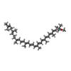


| #4: Chemical | ChemComp-LDA / Lauryldimethylamine oxide Mass: 229.402 Da / Num. of mol.: 5 / Source method: obtained synthetically / Formula: C14H31NO / Comment: LDAO, detergent*YM Mass: 229.402 Da / Num. of mol.: 5 / Source method: obtained synthetically / Formula: C14H31NO / Comment: LDAO, detergent*YM#5: Chemical | ChemComp-BCL / Bacteriochlorophyll Mass: 911.504 Da / Num. of mol.: 4 Mass: 911.504 Da / Num. of mol.: 4Source method: isolated from a genetically manipulated source Formula: C55H74MgN4O6 / Feature type: SUBJECT OF INVESTIGATION #6: Chemical | Pheophytin Mass: 889.215 Da / Num. of mol.: 2 Mass: 889.215 Da / Num. of mol.: 2Source method: isolated from a genetically manipulated source Formula: C55H76N4O6 / Feature type: SUBJECT OF INVESTIGATION #7: Chemical | Coenzyme Q10 Mass: 863.343 Da / Num. of mol.: 2 Mass: 863.343 Da / Num. of mol.: 2Source method: isolated from a genetically manipulated source Formula: C59H90O4 / Feature type: SUBJECT OF INVESTIGATION #8: Chemical | ChemComp-CL / | Chloride Mass: 35.453 Da / Num. of mol.: 1 / Source method: obtained synthetically / Formula: Cl Mass: 35.453 Da / Num. of mol.: 1 / Source method: obtained synthetically / Formula: Cl#9: Chemical | ChemComp-FE / | Iron Mass: 55.845 Da / Num. of mol.: 1 / Source method: isolated from a natural source / Formula: Fe Mass: 55.845 Da / Num. of mol.: 1 / Source method: isolated from a natural source / Formula: Fe#10: Chemical | ChemComp-SPO / |  Mass: 568.914 Da / Num. of mol.: 1 / Source method: isolated from a natural source / Formula: C41H60O Mass: 568.914 Da / Num. of mol.: 1 / Source method: isolated from a natural source / Formula: C41H60O#11: Chemical | ChemComp-CDL / | Cardiolipin Mass: 1464.043 Da / Num. of mol.: 1 / Source method: isolated from a natural source / Formula: C81H156O17P2 / Comment: phospholipid*YM Mass: 1464.043 Da / Num. of mol.: 1 / Source method: isolated from a natural source / Formula: C81H156O17P2 / Comment: phospholipid*YM#12: Water | ChemComp-HOH / | WaterMass: 18.015 Da / Num. of mol.: 244 / Source method: isolated from a natural source / Formula: H2O |
|---|
-Details
| Has ligand of interest | Y |
|---|
-Experimental details
-Experiment
| Experiment | Method: X-RAY DIFFRACTION / Number of used crystals: 1 |
|---|
- Sample preparation
| Crystal grow | Temperature: 293 K / Method: vapor diffusion, hanging drop / pH: 8 Details: 1 M potassium phosphate, 3.5% 1,2,3-heptanetriol, and 0.1% LDAO precipitant solution; 1.4-1.5 M potassium phosphate reservoir solution, pH 8.0, VAPOR DIFFUSION, HANGING DROP, temperature 293K |
|---|
-Data collection
| Diffraction | Mean temperature: 250 K / Serial crystal experiment: N | ||||||||||||||||||||||||||||||||||||||||||||||||||||||||||||||||||||||||||||||||||||||||||||||||||||||||||||||||||||||||||||||||||||||||||||||||||||||||||||||||||||||||||||||||||||||||||||||||||||||||||||||||||
|---|---|---|---|---|---|---|---|---|---|---|---|---|---|---|---|---|---|---|---|---|---|---|---|---|---|---|---|---|---|---|---|---|---|---|---|---|---|---|---|---|---|---|---|---|---|---|---|---|---|---|---|---|---|---|---|---|---|---|---|---|---|---|---|---|---|---|---|---|---|---|---|---|---|---|---|---|---|---|---|---|---|---|---|---|---|---|---|---|---|---|---|---|---|---|---|---|---|---|---|---|---|---|---|---|---|---|---|---|---|---|---|---|---|---|---|---|---|---|---|---|---|---|---|---|---|---|---|---|---|---|---|---|---|---|---|---|---|---|---|---|---|---|---|---|---|---|---|---|---|---|---|---|---|---|---|---|---|---|---|---|---|---|---|---|---|---|---|---|---|---|---|---|---|---|---|---|---|---|---|---|---|---|---|---|---|---|---|---|---|---|---|---|---|---|---|---|---|---|---|---|---|---|---|---|---|---|---|---|---|---|---|
| Diffraction source | Source: SYNCHROTRON / Site: SSRL / Beamline: BL9-2 / Wavelength: 0.97946 Å | ||||||||||||||||||||||||||||||||||||||||||||||||||||||||||||||||||||||||||||||||||||||||||||||||||||||||||||||||||||||||||||||||||||||||||||||||||||||||||||||||||||||||||||||||||||||||||||||||||||||||||||||||||
| Detector | Type: DECTRIS PILATUS 6M / Detector: PIXEL / Date: Aug 11, 2017 / Details: Rh coated collimating mirror, K-B focusing mirrors | ||||||||||||||||||||||||||||||||||||||||||||||||||||||||||||||||||||||||||||||||||||||||||||||||||||||||||||||||||||||||||||||||||||||||||||||||||||||||||||||||||||||||||||||||||||||||||||||||||||||||||||||||||
| Radiation | Protocol: SINGLE WAVELENGTH / Monochromatic (M) / Laue (L): M / Scattering type: x-ray | ||||||||||||||||||||||||||||||||||||||||||||||||||||||||||||||||||||||||||||||||||||||||||||||||||||||||||||||||||||||||||||||||||||||||||||||||||||||||||||||||||||||||||||||||||||||||||||||||||||||||||||||||||
| Radiation wavelength | Wavelength: 0.97946 Å / Relative weight: 1 | ||||||||||||||||||||||||||||||||||||||||||||||||||||||||||||||||||||||||||||||||||||||||||||||||||||||||||||||||||||||||||||||||||||||||||||||||||||||||||||||||||||||||||||||||||||||||||||||||||||||||||||||||||
| Reflection | Resolution: 2.48→39 Å / Num. obs: 76551 / % possible obs: 99.6 % / Redundancy: 5.076 % / Biso Wilson estimate: 58.55 Å2 / CC1/2: 0.996 / Rmerge(I) obs: 0.108 / Rrim(I) all: 0.121 / Χ2: 1.039 / Net I/σ(I): 10.16 / Num. measured all: 388566 / Scaling rejects: 6 | ||||||||||||||||||||||||||||||||||||||||||||||||||||||||||||||||||||||||||||||||||||||||||||||||||||||||||||||||||||||||||||||||||||||||||||||||||||||||||||||||||||||||||||||||||||||||||||||||||||||||||||||||||
| Reflection shell | Diffraction-ID: 1
|
- Processing
| Software |
| ||||||||||||||||||||||||||||||||||||||||||||||||||||||||||||||||||||||||||||||||||||||||||||||||||||
|---|---|---|---|---|---|---|---|---|---|---|---|---|---|---|---|---|---|---|---|---|---|---|---|---|---|---|---|---|---|---|---|---|---|---|---|---|---|---|---|---|---|---|---|---|---|---|---|---|---|---|---|---|---|---|---|---|---|---|---|---|---|---|---|---|---|---|---|---|---|---|---|---|---|---|---|---|---|---|---|---|---|---|---|---|---|---|---|---|---|---|---|---|---|---|---|---|---|---|---|---|---|
| Refinement | Method to determine structure: MOLECULAR REPLACEMENT Starting model: Model from the Chloro derivative Resolution: 2.48→39 Å / Cor.coef. Fo:Fc: 0.956 / Cor.coef. Fo:Fc free: 0.937 / WRfactor Rfree: 0.1826 / WRfactor Rwork: 0.1591 / FOM work R set: 0.8627 / SU B: 10.683 / SU ML: 0.127 / SU R Cruickshank DPI: 0.1736 / SU Rfree: 0.1542 / Cross valid method: THROUGHOUT / σ(F): 0 / ESU R: 0.174 / ESU R Free: 0.154 / Stereochemistry target values: MAXIMUM LIKELIHOOD Details: HYDROGENS HAVE BEEN ADDED IN THE RIDING POSITIONS U VALUES : WITH TLS ADDED
| ||||||||||||||||||||||||||||||||||||||||||||||||||||||||||||||||||||||||||||||||||||||||||||||||||||
| Solvent computation | Ion probe radii: 0.8 Å / Shrinkage radii: 0.8 Å / VDW probe radii: 1.2 Å / Solvent model: MASK | ||||||||||||||||||||||||||||||||||||||||||||||||||||||||||||||||||||||||||||||||||||||||||||||||||||
| Displacement parameters | Biso max: 128.23 Å2 / Biso mean: 48.26 Å2 / Biso min: 18.45 Å2
| ||||||||||||||||||||||||||||||||||||||||||||||||||||||||||||||||||||||||||||||||||||||||||||||||||||
| Refinement step | Cycle: final / Resolution: 2.48→39 Å
| ||||||||||||||||||||||||||||||||||||||||||||||||||||||||||||||||||||||||||||||||||||||||||||||||||||
| Refine LS restraints |
| ||||||||||||||||||||||||||||||||||||||||||||||||||||||||||||||||||||||||||||||||||||||||||||||||||||
| LS refinement shell | Resolution: 2.48→2.544 Å / Rfactor Rfree error: 0 / Total num. of bins used: 20
| ||||||||||||||||||||||||||||||||||||||||||||||||||||||||||||||||||||||||||||||||||||||||||||||||||||
| Refinement TLS params. | Method: refined / Refine-ID: X-RAY DIFFRACTION
| ||||||||||||||||||||||||||||||||||||||||||||||||||||||||||||||||||||||||||||||||||||||||||||||||||||
| Refinement TLS group |
|
