 Movie
Movie Controller

[English] 日本語
 Yorodumi
- PDB-7m6u: Crystal structure of a circular permutation and computationally d... -
+ Open data

- Basic information
| Entry | Database: PDB / ID: 7m6u | ||||||
|---|---|---|---|---|---|---|---|
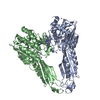
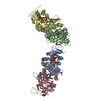
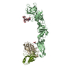
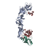
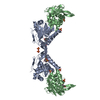
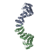
| Title | Crystal structure of a circular permutation and computationally designed pro-enzyme of carboxypeptidase G2 | ||||||
 Components Components | Carboxypeptidase G2 circular permuation pro-domain fusion | ||||||
 Keywords Keywords |  HYDROLASE / pro-enzyme / enzyme design / directed enzyme-prodrug therapy / methotrexate / circular permutation HYDROLASE / pro-enzyme / enzyme design / directed enzyme-prodrug therapy / methotrexate / circular permutation | ||||||
| Function / homology |  Function and homology informationglutamate carboxypeptidase / carboxypeptidase activity / metallopeptidase activity / proteolysis / metal ion binding Function and homology informationglutamate carboxypeptidase / carboxypeptidase activity / metallopeptidase activity / proteolysis / metal ion bindingSimilarity search - Function | ||||||
| Biological species |  Pseudomonas sp. (bacteria) Pseudomonas sp. (bacteria)synthetic construct (others) | ||||||
| Method | X-RAY DIFFRACTION / MOLECULAR REPLACEMENT / Resolution: 2.59 Å | ||||||
 Authors Authors | Yachnin, B.J. / Khare, S.D. | ||||||
| Funding support |  United States, 1items United States, 1items
| ||||||
 Citation Citation | Journal: Proc.Natl.Acad.Sci.USA / Year: 2022 Title: Massively parallel, computationally guided design of a proenzyme. Authors: Yachnin, B.J. / Azouz, L.R. / White 3rd, R.E. / Minetti, C.A.S.A. / Remeta, D.P. / Tan, V.M. / Drake, J.M. / Khare, S.D. #1: Journal: Biorxiv / Year: 2021Title: Massively parallel, computationally-guided design of a pro-enzyme Authors: Yachnin, B.J. / Azouz, L.R. / White, R.E. / Minetti, C.A.S.A. / Remeta, D.P. / Tan, V.M. / Drake, J.M. / Khare, S.D. | ||||||
| History |
|
- Structure visualization
| Structure viewer | Molecule: MolmilJmol/JSmol |
|---|
- Downloads & links
-Download
| PDBx/mmCIF format | 7m6u.cif.gz | 298.7 KB | Display | PDBx/mmCIF format |
|---|---|---|---|---|
| PDB format | pdb7m6u.ent.gz | 230.5 KB | Display | PDB format |
| PDBx/mmJSON format | 7m6u.json.gz | Tree view | PDBx/mmJSON format | |
| Others |  Other downloads Other downloads |
-Validation report
| Arichive directory | https://data.pdbj.org/pub/pdb/validation_reports/m6/7m6uftp://data.pdbj.org/pub/pdb/validation_reports/m6/7m6u | HTTPS FTP |
|---|
-Related structure data
| Related structure data |  1cg2S S: Starting model for refinement |
|---|---|
| Similar structure data | |
| Other databases |
|


-Links
 PDBj
PDBj

- Assembly
| Deposited unit | 
| ||||||||
|---|---|---|---|---|---|---|---|---|---|
| 1 | 
| ||||||||
| 2 | 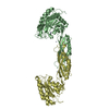
| ||||||||
| Unit cell |
|
-Components
| #1: Protein | Mass: 47217.277 Da / Num. of mol.: 4 / Mutation: K177A Source method: isolated from a genetically manipulated source Details: Pro-enzyme form Source: (gene. exp.) Pseudomonas sp. (strain RS-16) (bacteria), (gene. exp.) synthetic construct (others)Strain: RS-16 / Gene: cpg2 / Plasmid: pET15b / Production host: Escherichia coli BL21(DE3) (bacteria) / Strain (production host): BL21(DE3) / References: UniProt: P06621, glutamate carboxypeptidase#2: Chemical | ChemComp-ZN /   Mass: 65.409 Da / Num. of mol.: 20 / Source method: obtained synthetically / Formula: Zn Mass: 65.409 Da / Num. of mol.: 20 / Source method: obtained synthetically / Formula: Zn#3: Chemical | ChemComp-SO4 / | Sulfate  Mass: 96.063 Da / Num. of mol.: 1 / Source method: obtained synthetically / Formula: SO4 Mass: 96.063 Da / Num. of mol.: 1 / Source method: obtained synthetically / Formula: SO4#4: Water | ChemComp-HOH / | Water Mass: 18.015 Da / Num. of mol.: 132 / Source method: isolated from a natural source / Formula: H2O Mass: 18.015 Da / Num. of mol.: 132 / Source method: isolated from a natural source / Formula: H2OHas ligand of interest | N | |
|---|
-Experimental details
-Experiment
| Experiment | Method: X-RAY DIFFRACTION / Number of used crystals: 1 |
|---|
- Sample preparation
| Crystal | Density Matthews: 2.57 Å3/Da / Density % sol: 52.18 % |
|---|---|
| Crystal grow | Temperature: 293 K / Method: vapor diffusion, hanging drop / pH: 8 Details: Protein was prepared to a concentration of 19 mg/mL in 50 mM Tris 100 mM NaCl pH 7.4 0.2 mM ZnSO4. Reservoirs containing 750 uL of 20 mM Tris pH 8.0, 10% glycerol, and 10% PEG 3350 were ...Details: Protein was prepared to a concentration of 19 mg/mL in 50 mM Tris 100 mM NaCl pH 7.4 0.2 mM ZnSO4. Reservoirs containing 750 uL of 20 mM Tris pH 8.0, 10% glycerol, and 10% PEG 3350 were prepared in 24 well hanging drop vapor diffusion plates. Equal volumes of protein solution and reservoir solution were mixed on a cover slip and suspended over the reservoir |
-Data collection
| Diffraction | Mean temperature: 93 K / Serial crystal experiment: N | |||||||||||||||||||||||||||||||||||||||||||||||||||||||||||||||||||||||||||||||||||||||||||||||||||||||||||||||||||||||||||||||||||||||||||||||||||||||||||||||||||||||||||||||||||||||||||||
|---|---|---|---|---|---|---|---|---|---|---|---|---|---|---|---|---|---|---|---|---|---|---|---|---|---|---|---|---|---|---|---|---|---|---|---|---|---|---|---|---|---|---|---|---|---|---|---|---|---|---|---|---|---|---|---|---|---|---|---|---|---|---|---|---|---|---|---|---|---|---|---|---|---|---|---|---|---|---|---|---|---|---|---|---|---|---|---|---|---|---|---|---|---|---|---|---|---|---|---|---|---|---|---|---|---|---|---|---|---|---|---|---|---|---|---|---|---|---|---|---|---|---|---|---|---|---|---|---|---|---|---|---|---|---|---|---|---|---|---|---|---|---|---|---|---|---|---|---|---|---|---|---|---|---|---|---|---|---|---|---|---|---|---|---|---|---|---|---|---|---|---|---|---|---|---|---|---|---|---|---|---|---|---|---|---|---|---|---|---|---|
| Diffraction source | Source: ROTATING ANODE / Type: RIGAKU MICROMAX-007 / Wavelength: 1.54178 Å | |||||||||||||||||||||||||||||||||||||||||||||||||||||||||||||||||||||||||||||||||||||||||||||||||||||||||||||||||||||||||||||||||||||||||||||||||||||||||||||||||||||||||||||||||||||||||||||
| Detector | Type: RIGAKU RAXIS IV++ / Detector: IMAGE PLATE / Date: Jun 6, 2017 | |||||||||||||||||||||||||||||||||||||||||||||||||||||||||||||||||||||||||||||||||||||||||||||||||||||||||||||||||||||||||||||||||||||||||||||||||||||||||||||||||||||||||||||||||||||||||||||
| Radiation | Protocol: SINGLE WAVELENGTH / Monochromatic (M) / Laue (L): M / Scattering type: x-ray | |||||||||||||||||||||||||||||||||||||||||||||||||||||||||||||||||||||||||||||||||||||||||||||||||||||||||||||||||||||||||||||||||||||||||||||||||||||||||||||||||||||||||||||||||||||||||||||
| Radiation wavelength | Wavelength: 1.54178 Å / Relative weight: 1 | |||||||||||||||||||||||||||||||||||||||||||||||||||||||||||||||||||||||||||||||||||||||||||||||||||||||||||||||||||||||||||||||||||||||||||||||||||||||||||||||||||||||||||||||||||||||||||||
| Reflection | Resolution: 2.59→39.27 Å / Num. obs: 57919 / % possible obs: 97.2 % / Redundancy: 2.9 % / CC1/2: 0.952 / Rmerge(I) obs: 0.102 / Rpim(I) all: 0.071 / Rrim(I) all: 0.125 / Net I/σ(I): 6.8 | |||||||||||||||||||||||||||||||||||||||||||||||||||||||||||||||||||||||||||||||||||||||||||||||||||||||||||||||||||||||||||||||||||||||||||||||||||||||||||||||||||||||||||||||||||||||||||||
| Reflection shell | Diffraction-ID: 1
|
- Processing
| Software |
| ||||||||||||||||||||
|---|---|---|---|---|---|---|---|---|---|---|---|---|---|---|---|---|---|---|---|---|---|
| Refinement | Method to determine structure: MOLECULAR REPLACEMENT Starting model: 1CG2 Resolution: 2.59→39.27 Å / SU ML: 0.329 / Cross valid method: THROUGHOUT / ESU R: 1.72 / ESU R Free: 0.388 Details: Partway through the refinement, reflections between 4 and 3.4 A were deleted due to low data quality. Refining using the truncated dataset resulted in an improvement in Rfree by ~0.05, and ...Details: Partway through the refinement, reflections between 4 and 3.4 A were deleted due to low data quality. Refining using the truncated dataset resulted in an improvement in Rfree by ~0.05, and so the truncated dataset was used for the rest of refinement.
| ||||||||||||||||||||
| Solvent computation | Ion probe radii: 0.8 Å / Shrinkage radii: 0.8 Å / VDW probe radii: 1.2 Å | ||||||||||||||||||||
| Displacement parameters | Biso mean: 53.598 Å2
| ||||||||||||||||||||
| Refinement step | Cycle: LAST / Resolution: 2.59→39.27 Å
| ||||||||||||||||||||
| LS refinement shell | Resolution: 2.59→2.657 Å
|
