 Movie
Movie Controller


+ Open data

- Basic information
| Entry | Database: PDB / ID: 7lt8 | ||||||
|---|---|---|---|---|---|---|---|
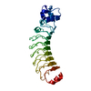
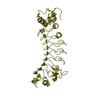
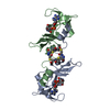
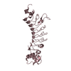
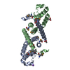
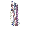
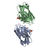
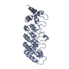
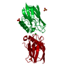
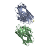
| Title | Crystal structure of Ras suppressor-1 | ||||||
 Components Components | Ras suppressor protein 1 | ||||||
 Keywords Keywords |  CELL ADHESION / leucine-rich repeat CELL ADHESION / leucine-rich repeat | ||||||
| Function / homology |  Function and homology information Function and homology informationRegulation of cytoskeletal remodeling and cell spreading by IPP complex components / positive regulation of cell-substrate adhesion / positive regulation of GTPase activity / focal adhesion / signal transduction / extracellular exosome / cytosolSimilarity search - Function | ||||||
| Biological species |  Homo sapiens (human) Homo sapiens (human) | ||||||
| Method | X-RAY DIFFRACTION / SYNCHROTRON / SAD / Resolution: 1.76000247097 Å | ||||||
 Authors Authors | Fukuda, K. / Qin, J. | ||||||
| Funding support |  United States, 1items United States, 1items
| ||||||
 Citation Citation | Journal: J.Biol.Chem. / Year: 2021 Title: Molecular basis for Ras suppressor-1 binding to PINCH-1 in focal adhesion assembly. Authors: Fukuda, K. / Lu, F. / Qin, J. | ||||||
| History |
|
- Structure visualization
| Structure viewer | Molecule: MolmilJmol/JSmol |
|---|
- Downloads & links
-Download
| PDBx/mmCIF format | 7lt8.cif.gz | 79 KB | Display | PDBx/mmCIF format |
|---|---|---|---|---|
| PDB format | pdb7lt8.ent.gz | 47 KB | Display | PDB format |
| PDBx/mmJSON format | 7lt8.json.gz | Tree view | PDBx/mmJSON format | |
| Others |  Other downloads Other downloads |
-Validation report
| Arichive directory | https://data.pdbj.org/pub/pdb/validation_reports/lt/7lt8ftp://data.pdbj.org/pub/pdb/validation_reports/lt/7lt8 | HTTPS FTP |
|---|
-Related structure data

-Links
 PDBj
PDBj


- Assembly
| Deposited unit | 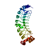
| ||||||||||
|---|---|---|---|---|---|---|---|---|---|---|---|
| 1 |
| ||||||||||
| Unit cell |
|
-Components
| #1: Protein | Mass: 31861.537 Da / Num. of mol.: 1 / Fragment: Leucine-rich repeat domain Source method: isolated from a genetically manipulated source Source: (gene. exp.) Homo sapiens (human) / Gene: RSU1, RSP1 / Production host:   Spodoptera frugiperda (fall armyworm) / Strain (production host): Sf-9 / References: UniProt: Q15404 Spodoptera frugiperda (fall armyworm) / Strain (production host): Sf-9 / References: UniProt: Q15404 |
|---|---|
| #2: Water | ChemComp-HOH / Water Mass: 18.015 Da / Num. of mol.: 131 / Source method: isolated from a natural source / Formula: H2O Mass: 18.015 Da / Num. of mol.: 131 / Source method: isolated from a natural source / Formula: H2O |
-Experimental details
-Experiment
| Experiment | Method: X-RAY DIFFRACTION / Number of used crystals: 1 |
|---|
- Sample preparation
| Crystal | Density Matthews: 1.86 Å3/Da / Density % sol: 33.81 % |
|---|---|
| Crystal grow | Temperature: 296 K / Method: vapor diffusion / pH: 8.5 / Details: PEG4000, lithium chloride |
-Data collection
| Diffraction | Mean temperature: 100 K / Serial crystal experiment: N |
|---|---|
| Diffraction source | Source: SYNCHROTRON / Site: APS / Beamline: 19-BM / Wavelength: 0.97919 Å |
| Detector | Type: ADSC QUANTUM 210r / Detector: CCD / Date: Aug 17, 2017 |
| Radiation | Protocol: SINGLE WAVELENGTH / Monochromatic (M) / Laue (L): M / Scattering type: x-ray |
| Radiation wavelength | Wavelength: 0.97919 Å / Relative weight: 1 |
| Reflection | Resolution: 1.76→50 Å / Num. obs: 22975 / % possible obs: 97.1 % / Redundancy: 4.8 % / Biso Wilson estimate: 22.2947677536 Å2 / Rmerge(I) obs: 0.084 / Net I/σ(I): 36.52 |
| Reflection shell | Resolution: 1.76→1.79 Å / Rmerge(I) obs: 0.804 / Num. unique obs: 1136 |
-Phasing
| Phasing | Method: SAD |
|---|
- Processing
| Software |
| ||||||||||||||||||||||||||||||||||||||||||||||||||||||||
|---|---|---|---|---|---|---|---|---|---|---|---|---|---|---|---|---|---|---|---|---|---|---|---|---|---|---|---|---|---|---|---|---|---|---|---|---|---|---|---|---|---|---|---|---|---|---|---|---|---|---|---|---|---|---|---|---|---|
| Refinement | Method to determine structure: SAD / Resolution: 1.76000247097→27.3413475696 Å / SU ML: 0.161477691195 / Cross valid method: THROUGHOUT / σ(F): 1.34364813708 / Phase error: 22.0795682201 Stereochemistry target values: GeoStd + Monomer Library + CDL v1.2
| ||||||||||||||||||||||||||||||||||||||||||||||||||||||||
| Solvent computation | Shrinkage radii: 0.9 Å / VDW probe radii: 1.11 Å / Solvent model: FLAT BULK SOLVENT MODEL | ||||||||||||||||||||||||||||||||||||||||||||||||||||||||
| Displacement parameters | Biso mean: 26.430761098 Å2 | ||||||||||||||||||||||||||||||||||||||||||||||||||||||||
| Refinement step | Cycle: LAST / Resolution: 1.76000247097→27.3413475696 Å
| ||||||||||||||||||||||||||||||||||||||||||||||||||||||||
| Refine LS restraints |
| ||||||||||||||||||||||||||||||||||||||||||||||||||||||||
| LS refinement shell |
|
