 Movie
Movie Controller

[English] 日本語
 Yorodumi
- PDB-6v3v: Assembly of VIQKI I456(beta-L-homoisoleucine)with human parainflu... -
+ Open data

- Basic information
| Entry | Database: PDB / ID: 6v3v | |||||||||
|---|---|---|---|---|---|---|---|---|---|---|
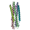
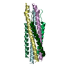
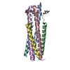
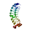
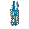
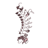
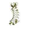
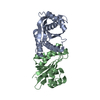
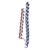
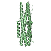
| Title | Assembly of VIQKI I456(beta-L-homoisoleucine)with human parainfluenza virus type 3 (HPIV3) fusion glycoprotein N-terminal heptad repeat domain | |||||||||
 Components Components | (Fusion glycoprotein F1) x 2 | |||||||||
 Keywords Keywords |  VIRAL PROTEIN / Fusion glycoprotein / six-helix bundle / foldamer VIRAL PROTEIN / Fusion glycoprotein / six-helix bundle / foldamer | |||||||||
| Function / homology | Precursor fusion glycoprotein F0, Paramyxoviridae / Fusion glycoprotein F0 / symbiont entry into host cell / fusion of virus membrane with host plasma membrane / viral envelope / host cell plasma membrane / virion membrane / membrane / Fusion glycoprotein F0 Function and homology information Function and homology information | |||||||||
| Biological species |  Human parainfluenza 3 virus Human parainfluenza 3 virus | |||||||||
| Method | X-RAY DIFFRACTION / SYNCHROTRON / MOLECULAR REPLACEMENT / Resolution: 2.17 Å | |||||||||
 Authors Authors | Outlaw, V.K. / Gellman, S.H. | |||||||||
| Funding support |  United States, 2items United States, 2items
| |||||||||
 Citation Citation | Journal: Acs Infect Dis. / Year: 2020 Title: Effects of Single alpha-to-beta Residue Replacements on Recognition of an Extended Segment in a Viral Fusion Protein. Authors: Outlaw, V.K. / Kreitler, D.F. / Stelitano, D. / Porotto, M. / Moscona, A. / Gellman, S.H. | |||||||||
| History |
|
- Structure visualization
| Structure viewer | Molecule: MolmilJmol/JSmol |
|---|
- Downloads & links
-Download
| PDBx/mmCIF format | 6v3v.cif.gz | 130.1 KB | Display | PDBx/mmCIF format |
|---|---|---|---|---|
| PDB format | pdb6v3v.ent.gz | 85.4 KB | Display | PDB format |
| PDBx/mmJSON format | 6v3v.json.gz | Tree view | PDBx/mmJSON format | |
| Others |  Other downloads Other downloads |
-Validation report
| Arichive directory | https://data.pdbj.org/pub/pdb/validation_reports/v3/6v3vftp://data.pdbj.org/pub/pdb/validation_reports/v3/6v3v | HTTPS FTP |
|---|
-Related structure data
| Related structure data |  6prlC  6pyqC  6pz6C 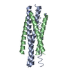 6vasC  6nroS S: Starting model for refinement C: citing same article ( |
|---|---|
| Similar structure data |
-Links
 PDBj
PDBj

- Assembly
| Deposited unit | 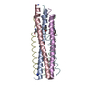
| ||||||||||||
|---|---|---|---|---|---|---|---|---|---|---|---|---|---|
| 1 |
| ||||||||||||
| Unit cell |
|
-Components
| #1: Protein | Mass: 5656.473 Da / Num. of mol.: 3 / Source method: obtained synthetically Source: (synth.) Human parainfluenza 3 virus (strain Wash/47885/57)References: UniProt: P06828 #2: Protein/peptide | Mass: 4208.922 Da / Num. of mol.: 3 Mutation: I456(beta-L-homoisoleucine), E459V, A463I, D466Q, Q479K, K480I Source method: obtained synthetically Source: (synth.) Human parainfluenza 3 virus (strain Wash/47885/57)References: UniProt: P06828 #3: Water | ChemComp-HOH / | Water Mass: 18.015 Da / Num. of mol.: 10 / Source method: isolated from a natural source / Formula: H2O Mass: 18.015 Da / Num. of mol.: 10 / Source method: isolated from a natural source / Formula: H2OHas ligand of interest | N | |
|---|
-Experimental details
-Experiment
| Experiment | Method: X-RAY DIFFRACTION / Number of used crystals: 1 |
|---|
- Sample preparation
| Crystal | Density Matthews: 1.94 Å3/Da / Density % sol: 36.74 % |
|---|---|
| Crystal grow | Temperature: 298 K / Method: vapor diffusion, hanging drop / pH: 7.5 Details: 30 mM sodium nitrate, 30 mM dibasic sodium phosphate, 30 mM ammonium sulfate, 100 mM HEPES/MOPS buffer (pH 7.5), 12.5% PEG1000, 12.5% PEG3350, 12.5% 2-methyl-2,4-pentanediol |
-Data collection
| Diffraction | Mean temperature: 100 K / Serial crystal experiment: N |
|---|---|
| Diffraction source | Source: SYNCHROTRON / Site: APS / Beamline: 21-ID-G / Wavelength: 0.97856 Å |
| Detector | Type: MARMOSAIC 300 mm CCD / Detector: CCD / Date: Aug 2, 2019 |
| Radiation | Protocol: SINGLE WAVELENGTH / Monochromatic (M) / Laue (L): M / Scattering type: x-ray |
| Radiation wavelength | Wavelength: 0.97856 Å / Relative weight: 1 |
| Reflection | Resolution: 2.17→28.02 Å / Num. obs: 12097 / % possible obs: 99.5 % / Redundancy: 7.5 % / Biso Wilson estimate: 52.31 Å2 / CC1/2: 1 / Rpim(I) all: 0.026 / Rrim(I) all: 0.072 / Rsym value: 0.067 / Net I/σ(I): 15.31 |
| Reflection shell | Resolution: 2.17→2.248 Å / Redundancy: 7.5 % / Mean I/σ(I) obs: 1.17 / Num. unique obs: 1195 / CC1/2: 0.521 / Rpim(I) all: 0.7275 / Rrim(I) all: 2.008 / Rsym value: 1.87 / % possible all: 99.5 |
- Processing
| Software |
| ||||||||||||||||||||||||||||||||||||||||||||||||||||||||||||||||||||||
|---|---|---|---|---|---|---|---|---|---|---|---|---|---|---|---|---|---|---|---|---|---|---|---|---|---|---|---|---|---|---|---|---|---|---|---|---|---|---|---|---|---|---|---|---|---|---|---|---|---|---|---|---|---|---|---|---|---|---|---|---|---|---|---|---|---|---|---|---|---|---|---|
| Refinement | Method to determine structure: MOLECULAR REPLACEMENT Starting model: 6NRO Resolution: 2.17→28.02 Å / SU ML: 0.355 / Cross valid method: FREE R-VALUE / σ(F): 1.35 / Phase error: 34.1648
| ||||||||||||||||||||||||||||||||||||||||||||||||||||||||||||||||||||||
| Solvent computation | Shrinkage radii: 0.9 Å / VDW probe radii: 1.11 Å | ||||||||||||||||||||||||||||||||||||||||||||||||||||||||||||||||||||||
| Displacement parameters | Biso mean: 76.63 Å2 | ||||||||||||||||||||||||||||||||||||||||||||||||||||||||||||||||||||||
| Refinement step | Cycle: LAST / Resolution: 2.17→28.02 Å
| ||||||||||||||||||||||||||||||||||||||||||||||||||||||||||||||||||||||
| Refine LS restraints |
| ||||||||||||||||||||||||||||||||||||||||||||||||||||||||||||||||||||||
| LS refinement shell |
| ||||||||||||||||||||||||||||||||||||||||||||||||||||||||||||||||||||||
| Refinement TLS params. | Method: refined / Origin x: -7.73623289111 Å / Origin y: -6.0967237848 Å / Origin z: -6.92619532569 Å
| ||||||||||||||||||||||||||||||||||||||||||||||||||||||||||||||||||||||
| Refinement TLS group | Selection details: all |
