 Movie
Movie Controller

[English] 日本語
 Yorodumi
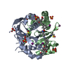
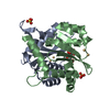
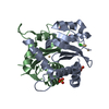
- PDB-7lqp: Rapid development of potent inhibitors of the HIV integrase-LEDGF... -
+ Open data

- Basic information
| Entry | Database: PDB / ID: 7lqp | ||||||
|---|---|---|---|---|---|---|---|
| Title | Rapid development of potent inhibitors of the HIV integrase-LEDGF interaction by fragment-linking using off-rate screening | ||||||
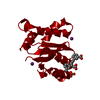
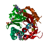
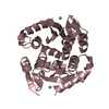
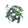
 Components Components | Integrase | ||||||
 Keywords Keywords | TRANSFERASE/INHIBITOR / Inhibitor / HIV Integrase / TRANSFERASE-INHIBITOR complex | ||||||
| Function / homology |  Function and homology information Function and homology informationRNA stem-loop binding / DNA integration / RNA-directed DNA polymerase activity / endonuclease activity / DNA recombination / symbiont entry into host cell / zinc ion bindingSimilarity search - Function | ||||||
| Biological species |   Human immunodeficiency virus 1 Human immunodeficiency virus 1 | ||||||
| Method | X-RAY DIFFRACTION / SYNCHROTRON / FOURIER SYNTHESIS / Resolution: 2.07 Å | ||||||
 Authors Authors | Gorman, M.A. / Parker, M.W. | ||||||
 Citation Citation | Journal: To Be Published Title: Rapid development of potent inhibitors of the HIV integrase-LEDGF interaction by fragment-linking using off-rate screening Authors: Gorman, M.A. / Parker, M.W. | ||||||
| History |
|
- Structure visualization
| Structure viewer | Molecule: MolmilJmol/JSmol |
|---|
- Downloads & links
-Download
| PDBx/mmCIF format | 7lqp.cif.gz | 71.1 KB | Display | PDBx/mmCIF format |
|---|---|---|---|---|
| PDB format | pdb7lqp.ent.gz | 55.8 KB | Display | PDB format |
| PDBx/mmJSON format | 7lqp.json.gz | Tree view | PDBx/mmJSON format | |
| Others |  Other downloads Other downloads |
-Validation report
| Arichive directory | https://data.pdbj.org/pub/pdb/validation_reports/lq/7lqpftp://data.pdbj.org/pub/pdb/validation_reports/lq/7lqp | HTTPS FTP |
|---|
-Related structure data


-Links
 PDBj
PDBj- Assembly
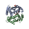
| Deposited unit | 
| ||||||||
|---|---|---|---|---|---|---|---|---|---|
| 1 |
| ||||||||
| Unit cell |
|
-Components
| #1: Protein | Mass: 17857.289 Da / Num. of mol.: 2 / Fragment: core domain (UNP residues 57-212) Source method: isolated from a genetically manipulated source Source: (gene. exp.) Human immunodeficiency virus 1 / Production host:  Escherichia coli (E. coli) / References: UniProt: Q76353 Escherichia coli (E. coli) / References: UniProt: Q76353#2: Chemical | ChemComp-IOD / Iodide  Mass: 126.904 Da / Num. of mol.: 6 / Source method: obtained synthetically / Formula: I Mass: 126.904 Da / Num. of mol.: 6 / Source method: obtained synthetically / Formula: I#3: Chemical | ChemComp-SO4 / Sulfate  Mass: 96.063 Da / Num. of mol.: 12 / Source method: obtained synthetically / Formula: SO4 Mass: 96.063 Da / Num. of mol.: 12 / Source method: obtained synthetically / Formula: SO4#4: Chemical | ChemComp-YAV / |   Mass: 780.817 Da / Num. of mol.: 1 / Source method: obtained synthetically / Formula: C46H40N2O10 / Feature type: SUBJECT OF INVESTIGATION Mass: 780.817 Da / Num. of mol.: 1 / Source method: obtained synthetically / Formula: C46H40N2O10 / Feature type: SUBJECT OF INVESTIGATION#5: Water | ChemComp-HOH / | Water Mass: 18.015 Da / Num. of mol.: 75 / Source method: isolated from a natural source / Formula: H2O Mass: 18.015 Da / Num. of mol.: 75 / Source method: isolated from a natural source / Formula: H2OHas ligand of interest | Y | |
|---|
-Experimental details
-Experiment
| Experiment | Method: X-RAY DIFFRACTION / Number of used crystals: 1 |
|---|
- Sample preparation
| Crystal | Density Matthews: 2.08 Å3/Da / Density % sol: 40.91 % / Description: Bi-pyramid |
|---|---|
| Crystal grow | Temperature: 293 K / Method: vapor diffusion, hanging drop / Details: 2.2 M ammonium sulfate, 100 mM sodium iodide |
-Data collection
| Diffraction | Mean temperature: 100 K / Serial crystal experiment: N |
|---|---|
| Diffraction source | Source: SYNCHROTRON / Site: Australian Synchrotron  / Beamline: MX2 / Wavelength: 0.9775 Å / Beamline: MX2 / Wavelength: 0.9775 Å |
| Detector | Type: ADSC QUANTUM 315r / Detector: CCD / Date: Aug 17, 2016 |
| Radiation | Protocol: SINGLE WAVELENGTH / Monochromatic (M) / Laue (L): M / Scattering type: x-ray |
| Radiation wavelength | Wavelength: 0.9775 Å / Relative weight: 1 |
| Reflection | Resolution: 2.07→46.42 Å / Num. obs: 17645 / % possible obs: 99.5 % / Redundancy: 5.5 % / Biso Wilson estimate: 23.2 Å2 / CC1/2: 0.992 / Rmerge(I) obs: 0.165 / Rpim(I) all: 0.078 / Net I/σ(I): 7.6 |
| Reflection shell | Resolution: 2.07→2.13 Å / Rmerge(I) obs: 0.726 / Mean I/σ(I) obs: 2.1 / Num. unique obs: 1300 / CC1/2: 0.613 / Rpim(I) all: 0.352 |
- Processing
| Software |
| |||||||||||||||||||||||||||||||||||||||||||||||||||||||||||||||||||||||||||||||||||||||||||||||||||||||||||||||||||||||||||||||||||||||||||||||||||
|---|---|---|---|---|---|---|---|---|---|---|---|---|---|---|---|---|---|---|---|---|---|---|---|---|---|---|---|---|---|---|---|---|---|---|---|---|---|---|---|---|---|---|---|---|---|---|---|---|---|---|---|---|---|---|---|---|---|---|---|---|---|---|---|---|---|---|---|---|---|---|---|---|---|---|---|---|---|---|---|---|---|---|---|---|---|---|---|---|---|---|---|---|---|---|---|---|---|---|---|---|---|---|---|---|---|---|---|---|---|---|---|---|---|---|---|---|---|---|---|---|---|---|---|---|---|---|---|---|---|---|---|---|---|---|---|---|---|---|---|---|---|---|---|---|---|---|---|---|
| Refinement | Method to determine structure: FOURIER SYNTHESIS / Resolution: 2.07→38.52 Å / Cross valid method: THROUGHOUT / σ(F): 13.15 / Phase error: 24.64 / Stereochemistry target values: TWIN_LSQ_F
| |||||||||||||||||||||||||||||||||||||||||||||||||||||||||||||||||||||||||||||||||||||||||||||||||||||||||||||||||||||||||||||||||||||||||||||||||||
| Solvent computation | Shrinkage radii: 0.9 Å / VDW probe radii: 1.11 Å / Solvent model: FLAT BULK SOLVENT MODEL | |||||||||||||||||||||||||||||||||||||||||||||||||||||||||||||||||||||||||||||||||||||||||||||||||||||||||||||||||||||||||||||||||||||||||||||||||||
| Refinement step | Cycle: LAST / Resolution: 2.07→38.52 Å
| |||||||||||||||||||||||||||||||||||||||||||||||||||||||||||||||||||||||||||||||||||||||||||||||||||||||||||||||||||||||||||||||||||||||||||||||||||
| Refine LS restraints |
| |||||||||||||||||||||||||||||||||||||||||||||||||||||||||||||||||||||||||||||||||||||||||||||||||||||||||||||||||||||||||||||||||||||||||||||||||||
| LS refinement shell |
|
