 Movie
Movie Controller

[English] 日本語
 Yorodumi
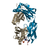
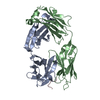
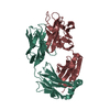
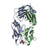
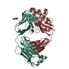
- PDB-7lkb: Crystal structure of PfCSP peptide 21 with vaccine-elicited human... -
+ Open data

- Basic information
| Entry | Database: PDB / ID: 7lkb | ||||||
|---|---|---|---|---|---|---|---|
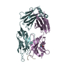
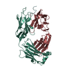
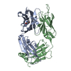
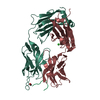
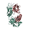
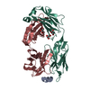
| Title | Crystal structure of PfCSP peptide 21 with vaccine-elicited human anti-malaria antibody m42.127 | ||||||
 Components Components |
| ||||||
 Keywords Keywords |  IMMUNE SYSTEM/ANTIGEN / Malaria / CSP / Junction region / antibody / m42.127 / IMMUNE SYSTEM / IMMUNE SYSTEM-ANTIGEN complex IMMUNE SYSTEM/ANTIGEN / Malaria / CSP / Junction region / antibody / m42.127 / IMMUNE SYSTEM / IMMUNE SYSTEM-ANTIGEN complex | ||||||
| Function / homology |  Function and homology information Function and homology informationentry into host cell by a symbiont-containing vacuole / side of membrane / cell surface / plasma membrane / cytoplasmSimilarity search - Function | ||||||
| Biological species |  Homo sapiens (human) Homo sapiens (human) Plasmodium falciparum 3D7 (eukaryote) Plasmodium falciparum 3D7 (eukaryote) | ||||||
| Method | X-RAY DIFFRACTION / SYNCHROTRON / MOLECULAR REPLACEMENT / Resolution: 1.8 Å | ||||||
 Authors Authors | Xu, K. / Kwong, P.D. | ||||||
 Citation Citation | Journal: Immunity / Year: 2021 Title: Vaccination in a humanized mouse model elicits highly protective PfCSP-targeting anti-malarial antibodies. Authors: Kratochvil, S. / Shen, C.H. / Lin, Y.C. / Xu, K. / Nair, U. / Da Silva Pereira, L. / Tripathi, P. / Arnold, J. / Chuang, G.Y. / Melzi, E. / Schon, A. / Zhang, B. / Dillon, M. / Bonilla, B. / ...Authors: Kratochvil, S. / Shen, C.H. / Lin, Y.C. / Xu, K. / Nair, U. / Da Silva Pereira, L. / Tripathi, P. / Arnold, J. / Chuang, G.Y. / Melzi, E. / Schon, A. / Zhang, B. / Dillon, M. / Bonilla, B. / Flynn, B.J. / Kirsch, K.H. / Kisalu, N.K. / Kiyuka, P.K. / Liu, T. / Ou, L. / Pancera, M. / Rawi, R. / Reveiz, M. / Seignon, K. / Wang, L.T. / Waring, M.T. / Warner, J. / Yang, Y. / Francica, J.R. / Idris, A.H. / Seder, R.A. / Kwong, P.D. / Batista, F.D. | ||||||
| History |
|
- Structure visualization
| Structure viewer | Molecule: MolmilJmol/JSmol |
|---|
- Downloads & links
-Download
| PDBx/mmCIF format | 7lkb.cif.gz | 202 KB | Display | PDBx/mmCIF format |
|---|---|---|---|---|
| PDB format | pdb7lkb.ent.gz | 156.9 KB | Display | PDB format |
| PDBx/mmJSON format | 7lkb.json.gz | Tree view | PDBx/mmJSON format | |
| Others |  Other downloads Other downloads |
-Validation report
| Arichive directory | https://data.pdbj.org/pub/pdb/validation_reports/lk/7lkbftp://data.pdbj.org/pub/pdb/validation_reports/lk/7lkb | HTTPS FTP |
|---|
-Related structure data
| Related structure data | 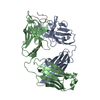 7lkgC  7rajC 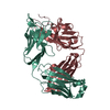 7rcsC 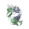 7rd3C 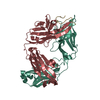 7rd4C  7rd9C 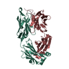 7rdaC 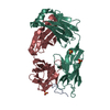 6b5mS S: Starting model for refinement C: citing same article ( |
|---|---|
| Similar structure data |


-Links
 PDBj
PDBj

- Assembly
| Deposited unit | 
| ||||||||
|---|---|---|---|---|---|---|---|---|---|
| 1 | 
| ||||||||
| 2 | 
| ||||||||
| Unit cell |
|
-Components
| #1: Antibody | Mass: 24996.148 Da / Num. of mol.: 2 Source method: isolated from a genetically manipulated source Source: (gene. exp.) Homo sapiens (human) / Production host: Homo sapiens (human)#2: Antibody | Mass: 24513.143 Da / Num. of mol.: 2 Source method: isolated from a genetically manipulated source Source: (gene. exp.) Homo sapiens (human) / Production host: Homo sapiens (human)#3: Protein/peptide | / CSMass: 1562.553 Da / Num. of mol.: 2 / Fragment: Peptide 21 / Source method: obtained synthetically / Source: (synth.) Plasmodium falciparum 3D7 (eukaryote) / References: UniProt: P02893#4: Chemical | ChemComp-SO4 / | Sulfate  Mass: 96.063 Da / Num. of mol.: 1 / Source method: obtained synthetically / Formula: SO4 Mass: 96.063 Da / Num. of mol.: 1 / Source method: obtained synthetically / Formula: SO4#5: Water | ChemComp-HOH / | Water Mass: 18.015 Da / Num. of mol.: 661 / Source method: isolated from a natural source / Formula: H2O Mass: 18.015 Da / Num. of mol.: 661 / Source method: isolated from a natural source / Formula: H2OHas ligand of interest | N | |
|---|
-Experimental details
-Experiment
| Experiment | Method: X-RAY DIFFRACTION / Number of used crystals: 1 |
|---|
- Sample preparation
| Crystal | Density Matthews: 2.18 Å3/Da / Density % sol: 43.66 % |
|---|---|
| Crystal grow | Temperature: 293 K / Method: vapor diffusion, sitting drop Details: 0.1 M Sodium acetate trihydrate pH 4.5, and 30% w/v polyethylene glycol 1500 |
-Data collection
| Diffraction | Mean temperature: 100 K / Serial crystal experiment: N | |||||||||||||||||||||||||||||||||||||||||||||||||||||||||||||||||||||||||||||||||||||||||||||||||||||||||||||||||||||||||||||||||||||||||||||||||||||||||||||||||||||||||||||||||||||||||||||
|---|---|---|---|---|---|---|---|---|---|---|---|---|---|---|---|---|---|---|---|---|---|---|---|---|---|---|---|---|---|---|---|---|---|---|---|---|---|---|---|---|---|---|---|---|---|---|---|---|---|---|---|---|---|---|---|---|---|---|---|---|---|---|---|---|---|---|---|---|---|---|---|---|---|---|---|---|---|---|---|---|---|---|---|---|---|---|---|---|---|---|---|---|---|---|---|---|---|---|---|---|---|---|---|---|---|---|---|---|---|---|---|---|---|---|---|---|---|---|---|---|---|---|---|---|---|---|---|---|---|---|---|---|---|---|---|---|---|---|---|---|---|---|---|---|---|---|---|---|---|---|---|---|---|---|---|---|---|---|---|---|---|---|---|---|---|---|---|---|---|---|---|---|---|---|---|---|---|---|---|---|---|---|---|---|---|---|---|---|---|---|
| Diffraction source | Source: SYNCHROTRON / Site: APS  / Beamline: 22-ID / Wavelength: 1 Å / Beamline: 22-ID / Wavelength: 1 Å | |||||||||||||||||||||||||||||||||||||||||||||||||||||||||||||||||||||||||||||||||||||||||||||||||||||||||||||||||||||||||||||||||||||||||||||||||||||||||||||||||||||||||||||||||||||||||||||
| Detector | Type: DECTRIS EIGER X 16M / Detector: PIXEL / Date: Aug 1, 2020 | |||||||||||||||||||||||||||||||||||||||||||||||||||||||||||||||||||||||||||||||||||||||||||||||||||||||||||||||||||||||||||||||||||||||||||||||||||||||||||||||||||||||||||||||||||||||||||||
| Radiation | Protocol: SINGLE WAVELENGTH / Monochromatic (M) / Laue (L): M / Scattering type: x-ray | |||||||||||||||||||||||||||||||||||||||||||||||||||||||||||||||||||||||||||||||||||||||||||||||||||||||||||||||||||||||||||||||||||||||||||||||||||||||||||||||||||||||||||||||||||||||||||||
| Radiation wavelength | Wavelength: 1 Å / Relative weight: 1 | |||||||||||||||||||||||||||||||||||||||||||||||||||||||||||||||||||||||||||||||||||||||||||||||||||||||||||||||||||||||||||||||||||||||||||||||||||||||||||||||||||||||||||||||||||||||||||||
| Reflection | Resolution: 1.8→50 Å / Num. obs: 70885 / % possible obs: 89.3 % / Redundancy: 2.1 % / Rmerge(I) obs: 0.041 / Rpim(I) all: 0.037 / Rrim(I) all: 0.055 / Χ2: 1.079 / Net I/σ(I): 12.2 / Num. measured all: 146677 | |||||||||||||||||||||||||||||||||||||||||||||||||||||||||||||||||||||||||||||||||||||||||||||||||||||||||||||||||||||||||||||||||||||||||||||||||||||||||||||||||||||||||||||||||||||||||||||
| Reflection shell | Diffraction-ID: 1
|
- Processing
| Software |
| ||||||||||||||||||||||||||||||||||||||||||||||||||||||||||||||||||||||||||||||||||||||||||||||||||||||||||||||||||||||||||||||||||||||||||||||||||||||||||||||||||||||||||||||||||||||
|---|---|---|---|---|---|---|---|---|---|---|---|---|---|---|---|---|---|---|---|---|---|---|---|---|---|---|---|---|---|---|---|---|---|---|---|---|---|---|---|---|---|---|---|---|---|---|---|---|---|---|---|---|---|---|---|---|---|---|---|---|---|---|---|---|---|---|---|---|---|---|---|---|---|---|---|---|---|---|---|---|---|---|---|---|---|---|---|---|---|---|---|---|---|---|---|---|---|---|---|---|---|---|---|---|---|---|---|---|---|---|---|---|---|---|---|---|---|---|---|---|---|---|---|---|---|---|---|---|---|---|---|---|---|---|---|---|---|---|---|---|---|---|---|---|---|---|---|---|---|---|---|---|---|---|---|---|---|---|---|---|---|---|---|---|---|---|---|---|---|---|---|---|---|---|---|---|---|---|---|---|---|---|---|
| Refinement | Method to determine structure: MOLECULAR REPLACEMENT Starting model: 6B5M Resolution: 1.8→36.7 Å / SU ML: 0.2 / Cross valid method: THROUGHOUT / σ(F): 2 / Phase error: 22.84 / Stereochemistry target values: ML
| ||||||||||||||||||||||||||||||||||||||||||||||||||||||||||||||||||||||||||||||||||||||||||||||||||||||||||||||||||||||||||||||||||||||||||||||||||||||||||||||||||||||||||||||||||||||
| Solvent computation | Shrinkage radii: 0.9 Å / VDW probe radii: 1.11 Å / Solvent model: FLAT BULK SOLVENT MODEL | ||||||||||||||||||||||||||||||||||||||||||||||||||||||||||||||||||||||||||||||||||||||||||||||||||||||||||||||||||||||||||||||||||||||||||||||||||||||||||||||||||||||||||||||||||||||
| Displacement parameters | Biso max: 86.72 Å2 / Biso mean: 26.7531 Å2 / Biso min: 11.74 Å2 | ||||||||||||||||||||||||||||||||||||||||||||||||||||||||||||||||||||||||||||||||||||||||||||||||||||||||||||||||||||||||||||||||||||||||||||||||||||||||||||||||||||||||||||||||||||||
| Refinement step | Cycle: final / Resolution: 1.8→36.7 Å
| ||||||||||||||||||||||||||||||||||||||||||||||||||||||||||||||||||||||||||||||||||||||||||||||||||||||||||||||||||||||||||||||||||||||||||||||||||||||||||||||||||||||||||||||||||||||
| Refine LS restraints |
| ||||||||||||||||||||||||||||||||||||||||||||||||||||||||||||||||||||||||||||||||||||||||||||||||||||||||||||||||||||||||||||||||||||||||||||||||||||||||||||||||||||||||||||||||||||||
| LS refinement shell | Refine-ID: X-RAY DIFFRACTION / Rfactor Rfree error: 0 / Total num. of bins used: 25
|
