 Movie
Movie Controller


+ Open data

- Basic information
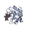
| Entry | Database: PDB / ID: 7l8q | |||||||||||||||||||||||||||
|---|---|---|---|---|---|---|---|---|---|---|---|---|---|---|---|---|---|---|---|---|---|---|---|---|---|---|---|---|
| Title | Crystal structure of human GPX4-U46C with oxidized Cys-46 | |||||||||||||||||||||||||||
 Components Components | Phospholipid hydroperoxide glutathione peroxidase | |||||||||||||||||||||||||||
 Keywords Keywords |  OXIDOREDUCTASE / alpha-beta protein OXIDOREDUCTASE / alpha-beta protein | |||||||||||||||||||||||||||
| Function / homology |  Function and homology informationphospholipid-hydroperoxide glutathione peroxidase / phospholipid-hydroperoxide glutathione peroxidase activity / Synthesis of 15-eicosatetraenoic acid derivatives / Synthesis of 12-eicosatetraenoic acid derivatives / negative regulation of ferroptosis / selenium binding / glutathione peroxidase / Synthesis of 5-eicosatetraenoic acids / lipoxygenase pathway / arachidonic acid metabolic process ...phospholipid-hydroperoxide glutathione peroxidase / phospholipid-hydroperoxide glutathione peroxidase activity / Synthesis of 15-eicosatetraenoic acid derivatives / Synthesis of 12-eicosatetraenoic acid derivatives / negative regulation of ferroptosis / selenium binding / glutathione peroxidase / Synthesis of 5-eicosatetraenoic acids / lipoxygenase pathway / arachidonic acid metabolic process / Biosynthesis of aspirin-triggered D-series resolvins / Biosynthesis of E-series 18(R)-resolvins / Biosynthesis of D-series resolvins / Biosynthesis of E-series 18(S)-resolvins / glutathione peroxidase activity / long-chain fatty acid biosynthetic process / protein polymerization / phospholipid metabolic process / response to estradiol / nuclear envelope / chromatin organization / cellular response to oxidative stress / spermatogenesis / response to oxidative stress / protein-containing complex / mitochondrion / extracellular exosome / identical protein binding / nucleus / cytosol Function and homology informationphospholipid-hydroperoxide glutathione peroxidase / phospholipid-hydroperoxide glutathione peroxidase activity / Synthesis of 15-eicosatetraenoic acid derivatives / Synthesis of 12-eicosatetraenoic acid derivatives / negative regulation of ferroptosis / selenium binding / glutathione peroxidase / Synthesis of 5-eicosatetraenoic acids / lipoxygenase pathway / arachidonic acid metabolic process ...phospholipid-hydroperoxide glutathione peroxidase / phospholipid-hydroperoxide glutathione peroxidase activity / Synthesis of 15-eicosatetraenoic acid derivatives / Synthesis of 12-eicosatetraenoic acid derivatives / negative regulation of ferroptosis / selenium binding / glutathione peroxidase / Synthesis of 5-eicosatetraenoic acids / lipoxygenase pathway / arachidonic acid metabolic process / Biosynthesis of aspirin-triggered D-series resolvins / Biosynthesis of E-series 18(R)-resolvins / Biosynthesis of D-series resolvins / Biosynthesis of E-series 18(S)-resolvins / glutathione peroxidase activity / long-chain fatty acid biosynthetic process / protein polymerization / phospholipid metabolic process / response to estradiol / nuclear envelope / chromatin organization / cellular response to oxidative stress / spermatogenesis / response to oxidative stress / protein-containing complex / mitochondrion / extracellular exosome / identical protein binding / nucleus / cytosolSimilarity search - Function | |||||||||||||||||||||||||||
| Biological species |  Homo sapiens (human) Homo sapiens (human) | |||||||||||||||||||||||||||
| Method | X-RAY DIFFRACTION / SYNCHROTRON / MOLECULAR REPLACEMENT / Resolution: 1.48 Å | |||||||||||||||||||||||||||
 Authors Authors | Forouhar, F. / Liu, H. / Seibt, T. / Saneto, R. / Wigby, K. / Friedman, J. / Xia, X. / Shchepinov, M.S. / Ramesh, S. / Conrad, M. / Stockwell, B.R. | |||||||||||||||||||||||||||
| Funding support |  United States, United States,  Germany, Germany,  Russian Federation, European Union, 8items Russian Federation, European Union, 8items
| |||||||||||||||||||||||||||
 Citation Citation | Journal: Nat.Chem.Biol. / Year: 2021 Title: Patient-derived variant of GPX4 reveals the structural basis for its catalytic activity and degradation mechanism Authors: Liu, H. / Forouhar, F. / Seibt, T. / Saneto, R. / Wigby, K. / Friedman, J. / Xia, X. / Shchepinov, M.S. / Ramesh, S. / Conrad, M. / Stockwell, B.R. | |||||||||||||||||||||||||||
| History |
|
- Structure visualization
| Structure viewer | Molecule: MolmilJmol/JSmol |
|---|
- Downloads & links
-Download
| PDBx/mmCIF format | 7l8q.cif.gz | 88.3 KB | Display | PDBx/mmCIF format |
|---|---|---|---|---|
| PDB format | pdb7l8q.ent.gz | 62.3 KB | Display | PDB format |
| PDBx/mmJSON format | 7l8q.json.gz | Tree view | PDBx/mmJSON format | |
| Others |  Other downloads Other downloads |
-Validation report
| Arichive directory | https://data.pdbj.org/pub/pdb/validation_reports/l8/7l8qftp://data.pdbj.org/pub/pdb/validation_reports/l8/7l8q | HTTPS FTP |
|---|
-Related structure data
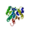
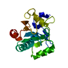
| Related structure data |  7l8kSC  7l8lC 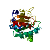 7l8mC  7l8rC S: Starting model for refinement C: citing same article ( |
|---|---|
| Similar structure data |


-Links
 PDBj
PDBj
- Assembly
| Deposited unit | 
| ||||||||
|---|---|---|---|---|---|---|---|---|---|
| 1 |
| ||||||||
| Unit cell |
|
-Components
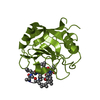
| #1: Protein | Mass: 21936.096 Da / Num. of mol.: 1 / Mutation: C46 is oxidized to sulfone Source method: isolated from a genetically manipulated source Details: Cys-46 is oxidized, so I named it SFO. / Source: (gene. exp.) Homo sapiens (human) / Gene: GPX4 / Production host:  Escherichia coli (E. coli) Escherichia coli (E. coli)References: UniProt: P36969, phospholipid-hydroperoxide glutathione peroxidase | ||||
|---|---|---|---|---|---|
| #2: Chemical | Acetate  Mass: 59.044 Da / Num. of mol.: 2 / Source method: obtained synthetically / Formula: C2H3O2 Mass: 59.044 Da / Num. of mol.: 2 / Source method: obtained synthetically / Formula: C2H3O2#3: Water | ChemComp-HOH / | Water Mass: 18.015 Da / Num. of mol.: 212 / Source method: isolated from a natural source / Formula: H2O Mass: 18.015 Da / Num. of mol.: 212 / Source method: isolated from a natural source / Formula: H2OHas ligand of interest | Y | |
-Experimental details
-Experiment
| Experiment | Method: X-RAY DIFFRACTION / Number of used crystals: 1 |
|---|
- Sample preparation
| Crystal | Density Matthews: 1.65 Å3/Da / Density % sol: 25.66 % |
|---|---|
| Crystal grow | Temperature: 293 K / Method: microbatch / pH: 5 Details: 0.1 M potassium thiocyanate, 0.1 M sodium acetate, and 20% (w/v) PEG 8000 |
-Data collection
| Diffraction | Mean temperature: 100 K / Serial crystal experiment: N |
|---|---|
| Diffraction source | Source: SYNCHROTRON / Site: APS / Beamline: 24-ID-E / Wavelength: 0.979 Å |
| Detector | Type: DECTRIS EIGER X 16M / Detector: PIXEL / Date: Nov 18, 2018 |
| Radiation | Protocol: SINGLE WAVELENGTH / Monochromatic (M) / Laue (L): M / Scattering type: x-ray |
| Radiation wavelength | Wavelength: 0.979 Å / Relative weight: 1 |
| Reflection | Resolution: 1.48→69.04 Å / Num. obs: 23698 / % possible obs: 99 % / Redundancy: 3.3 % / CC1/2: 0.99 / Net I/σ(I): 16 |
| Reflection shell | Resolution: 1.48→1.5 Å / Redundancy: 2.8 % / Num. unique obs: 1049 / CC1/2: 0.64 / % possible all: 87.4 |
- Processing
| Software |
| ||||||||||||||||||||||||||||||||||||||||||||||||||||||||||||||||||||||||||||||||||||||||||||||||||||||||||||||||||||||||||||||
|---|---|---|---|---|---|---|---|---|---|---|---|---|---|---|---|---|---|---|---|---|---|---|---|---|---|---|---|---|---|---|---|---|---|---|---|---|---|---|---|---|---|---|---|---|---|---|---|---|---|---|---|---|---|---|---|---|---|---|---|---|---|---|---|---|---|---|---|---|---|---|---|---|---|---|---|---|---|---|---|---|---|---|---|---|---|---|---|---|---|---|---|---|---|---|---|---|---|---|---|---|---|---|---|---|---|---|---|---|---|---|---|---|---|---|---|---|---|---|---|---|---|---|---|---|---|---|---|
| Refinement | Method to determine structure: MOLECULAR REPLACEMENT Starting model: 7L8K Resolution: 1.48→34.52 Å / SU ML: 0.2 / Cross valid method: THROUGHOUT / σ(F): 1.44 / Phase error: 20.2 / Stereochemistry target values: ML
| ||||||||||||||||||||||||||||||||||||||||||||||||||||||||||||||||||||||||||||||||||||||||||||||||||||||||||||||||||||||||||||||
| Solvent computation | Shrinkage radii: 0.9 Å / VDW probe radii: 1.11 Å / Solvent model: FLAT BULK SOLVENT MODEL | ||||||||||||||||||||||||||||||||||||||||||||||||||||||||||||||||||||||||||||||||||||||||||||||||||||||||||||||||||||||||||||||
| Displacement parameters | Biso max: 77.23 Å2 / Biso mean: 22.5835 Å2 / Biso min: 7.07 Å2 | ||||||||||||||||||||||||||||||||||||||||||||||||||||||||||||||||||||||||||||||||||||||||||||||||||||||||||||||||||||||||||||||
| Refinement step | Cycle: final / Resolution: 1.48→34.52 Å
| ||||||||||||||||||||||||||||||||||||||||||||||||||||||||||||||||||||||||||||||||||||||||||||||||||||||||||||||||||||||||||||||
| LS refinement shell | Refine-ID: X-RAY DIFFRACTION / Rfactor Rfree error: 0 / Total num. of bins used: 17
| ||||||||||||||||||||||||||||||||||||||||||||||||||||||||||||||||||||||||||||||||||||||||||||||||||||||||||||||||||||||||||||||
| Refinement TLS params. | Method: refined / Origin x: 0.1821 Å / Origin y: -0.3321 Å / Origin z: 13.1804 Å
| ||||||||||||||||||||||||||||||||||||||||||||||||||||||||||||||||||||||||||||||||||||||||||||||||||||||||||||||||||||||||||||||
| Refinement TLS group |
|
