[English] 日本語
Yorodumi
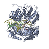
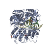
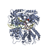
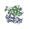
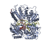
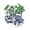
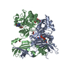
- PDB-7l35: Human DNA Ligase 1 - R771W nicked DNA complex -

+
Open data


ID or keywords:

Loading...

-
Basic information

Entry
Database: PDB / ID: 7l35
TitleHuman DNA Ligase 1 - R771W nicked DNA complex
Components
  • DNA (5'-D(*GP*CP*TP*GP*AP*TP*GP*CP*GP*TP*C)-3')
  • DNA (5'-D(*GP*TP*CP*CP*GP*AP*CP*GP*AP*CP*GP*CP*AP*TP*CP*AP*GP*C)-3')
  • DNA (5'-D(P*GP*TP*CP*GP*GP*AP*C)-3')
  • DNA ligase 1
KeywordsLIGASE/DNA / DNA ligase / LIGASE / LIGASE-DNA complex
Function / homology
Function and homology information


Okazaki fragment processing involved in mitotic DNA replication / DNA ligase activity / DNA ligase (ATP) / DNA ligase (ATP) activity / Processive synthesis on the lagging strand / DNA ligation / Processive synthesis on the C-strand of the telomere / Mismatch repair (MMR) directed by MSH2:MSH3 (MutSbeta) / Mismatch repair (MMR) directed by MSH2:MSH6 (MutSalpha) / lagging strand elongation ...Okazaki fragment processing involved in mitotic DNA replication / DNA ligase activity / DNA ligase (ATP) / DNA ligase (ATP) activity / Processive synthesis on the lagging strand / DNA ligation / Processive synthesis on the C-strand of the telomere / Mismatch repair (MMR) directed by MSH2:MSH3 (MutSbeta) / Mismatch repair (MMR) directed by MSH2:MSH6 (MutSalpha) / lagging strand elongation / DNA biosynthetic process / Early Phase of HIV Life Cycle / POLB-Dependent Long Patch Base Excision Repair / PCNA-Dependent Long Patch Base Excision Repair / anatomical structure morphogenesis / mismatch repair / base-excision repair, gap-filling / Gap-filling DNA repair synthesis and ligation in GG-NER / base-excision repair / Gap-filling DNA repair synthesis and ligation in TC-NER / DNA recombination / cell division / intracellular membrane-bounded organelle / DNA repair / mitochondrion / DNA binding / nucleoplasm / ATP binding / metal ion binding / nucleus
Similarity search - Function
DNA ligase, ATP-dependent / DNA ligase, ATP-dependent, N-terminal / DNA ligase, ATP-dependent, N-terminal domain superfamily / DNA ligase N terminus / ATP-dependent DNA ligase signature 2. / ATP-dependent DNA ligase AMP-binding site. / DNA ligase, ATP-dependent, C-terminal / ATP dependent DNA ligase C terminal region / DNA ligase, ATP-dependent, conserved site / ATP-dependent DNA ligase family profile. ...DNA ligase, ATP-dependent / DNA ligase, ATP-dependent, N-terminal / DNA ligase, ATP-dependent, N-terminal domain superfamily / DNA ligase N terminus / ATP-dependent DNA ligase signature 2. / ATP-dependent DNA ligase AMP-binding site. / DNA ligase, ATP-dependent, C-terminal / ATP dependent DNA ligase C terminal region / DNA ligase, ATP-dependent, conserved site / ATP-dependent DNA ligase family profile. / DNA ligase, ATP-dependent, central / ATP dependent DNA ligase domain / Nucleic acid-binding, OB-fold
Similarity search - Domain/homology
ADENOSINE MONOPHOSPHATE / DI(HYDROXYETHYL)ETHER / DNA / DNA (> 10) / DNA ligase 1
Similarity search - Component
Biological speciesHomo sapiens (human)
MethodX-RAY DIFFRACTION / SYNCHROTRON / MOLECULAR REPLACEMENT / Resolution: 2 Å
AuthorsTumbale, P.P. / Williams, R.S. / Schellenberg, M.S.
Funding support United States, 1items
OrganizationGrant numberCountry
National Institutes of Health/National Institute of Environmental Health Sciences (NIH/NIEHS)Z01ES102765 United States
CitationJournal: Nucleic Acids Res. / Year: 2021
Title: LIG1 syndrome mutations remodel a cooperative network of ligand binding interactions to compromise ligation efficiency.
Authors: Jurkiw, T.J. / Tumbale, P.P. / Schellenberg, M.J. / Cunningham-Rundles, C. / Williams, R.S. / O'Brien, P.J.
History
DepositionDec 17, 2020Deposition site: RCSB / Processing site: RCSB
Revision 1.0Jan 13, 2021Provider: repository / Type: Initial release
Revision 1.1Jan 27, 2021Group: Database references / Category: citation
Item: _citation.country / _citation.journal_abbrev ..._citation.country / _citation.journal_abbrev / _citation.journal_id_ASTM / _citation.journal_id_CSD / _citation.journal_id_ISSN / _citation.pdbx_database_id_DOI / _citation.pdbx_database_id_PubMed / _citation.title / _citation.year
Revision 1.2Mar 3, 2021Group: Database references / Category: citation
Item: _citation.journal_volume / _citation.page_first / _citation.page_last
Revision 1.3Oct 18, 2023Group: Data collection / Database references / Refinement description
Category: chem_comp_atom / chem_comp_bond ...chem_comp_atom / chem_comp_bond / database_2 / pdbx_initial_refinement_model
Item: _database_2.pdbx_DOI / _database_2.pdbx_database_accession

-
Structure visualization

Structure viewerMolecule:
MolmilJmol/JSmol

Downloads & links

-
Assembly

Deposited unit
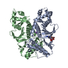
A: DNA ligase 1
B: DNA (5'-D(*GP*CP*TP*GP*AP*TP*GP*CP*GP*TP*C)-3')
C: DNA (5'-D(P*GP*TP*CP*GP*GP*AP*C)-3')
D: DNA (5'-D(*GP*TP*CP*CP*GP*AP*CP*GP*AP*CP*GP*CP*AP*TP*CP*AP*GP*C)-3')
hetero molecules


Theoretical massNumber of molelcules
Total (without water)83,6468
Polymers83,0474
Non-polymers5984
Water10,899605
1


  • Idetical with deposited unit
  • defined by author&software
TypeNameSymmetry operationNumber
identity operation1_555x,y,z1
Buried area8420 Å2
ΔGint-54 kcal/mol
Surface area30010 Å2
MethodPISA
Unit cell
Length a, b, c (Å)71.330, 100.523, 116.088
Angle α, β, γ (deg.)90.000, 90.000, 90.000
Int Tables number19
Space group name H-MP212121

-
Components

-
Protein , 1 types, 1 molecules A

#1: Protein DNA ligase 1 / / DNA ligase I / Polydeoxyribonucleotide synthase [ATP] 1


Mass: 72057.258 Da / Num. of mol.: 1 / Mutation: R771W
Source method: isolated from a genetically manipulated source
Source: (gene. exp.) Homo sapiens (human) / Gene: LIG1 / Production host: Escherichia coli (E. coli) / References: UniProt: P18858, DNA ligase (ATP)

-
DNA chain , 3 types, 3 molecules BCD

#2: DNA chain DNA (5'-D(*GP*CP*TP*GP*AP*TP*GP*CP*GP*TP*C)-3')


Mass: 3365.196 Da / Num. of mol.: 1 / Source method: obtained synthetically / Source: (synth.) Homo sapiens (human)
#3: DNA chain DNA (5'-D(P*GP*TP*CP*GP*GP*AP*C)-3')


Mass: 2138.423 Da / Num. of mol.: 1 / Source method: obtained synthetically / Source: (synth.) Homo sapiens (human)
#4: DNA chain DNA (5'-D(*GP*TP*CP*CP*GP*AP*CP*GP*AP*CP*GP*CP*AP*TP*CP*AP*GP*C)-3')


Mass: 5486.557 Da / Num. of mol.: 1 / Source method: obtained synthetically / Source: (synth.) Homo sapiens (human)

-
Non-polymers , 5 types, 609 molecules

#5: Chemical ChemComp-PEG / DI(HYDROXYETHYL)ETHER / Diethylene glycol


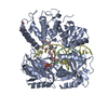
Mass: 106.120 Da / Num. of mol.: 1 / Source method: obtained synthetically / Formula: C4H10O3
#6: Chemical ChemComp-NA / SODIUM ION


Mass: 22.990 Da / Num. of mol.: 1 / Source method: obtained synthetically / Formula: Na
#7: Chemical ChemComp-AMP / ADENOSINE MONOPHOSPHATE / Adenosine monophosphate


Mass: 347.221 Da / Num. of mol.: 1 / Source method: obtained synthetically / Formula: C10H14N5O7P / Feature type: SUBJECT OF INVESTIGATION / Comment: AMP*YM
#8: Chemical ChemComp-TRS / 2-AMINO-2-HYDROXYMETHYL-PROPANE-1,3-DIOL / TRIS BUFFER / Tris


Mass: 122.143 Da / Num. of mol.: 1 / Source method: obtained synthetically / Formula: C4H12NO3 / Comment: pH buffer*YM
#9: Water ChemComp-HOH / water / Water


Mass: 18.015 Da / Num. of mol.: 605 / Source method: isolated from a natural source / Formula: H2O

-
Details

Has ligand of interestY

-
Experimental details

-
Experiment

ExperimentMethod: X-RAY DIFFRACTION / Number of used crystals: 1

-
Sample preparation

CrystalDensity Matthews: 2.5 Å3/Da / Density % sol: 50.77 %
Crystal growTemperature: 293 K / Method: vapor diffusion, hanging drop / pH: 6
Details: 100 mM MES, pH 6, 100 mM lithium acetate, 15% (w/v) polyethylene glycol PEG3350

-
Data collection

DiffractionMean temperature: 100 K / Serial crystal experiment: N
Diffraction sourceSource: SYNCHROTRON / Site: APS / Beamline: 22-ID / Wavelength: 1 Å
DetectorType: DECTRIS EIGER X 16M / Detector: PIXEL / Date: Feb 17, 2017
RadiationProtocol: SINGLE WAVELENGTH / Monochromatic (M) / Laue (L): M / Scattering type: x-ray
Radiation wavelengthWavelength: 1 Å / Relative weight: 1
ReflectionResolution: 2→50 Å / Num. obs: 57075 / % possible obs: 99.87 % / Redundancy: 7.4 % / CC1/2: 0.996 / Rmerge(I) obs: 0.11 / Net I/σ(I): 15.9
Reflection shellResolution: 2→2.07 Å / Rmerge(I) obs: 1.046 / Num. unique obs: 5634 / CC1/2: 0.763

-
Processing

Software
NameVersionClassification
PHENIX1.10.1_2155refinement
PDB_EXTRACT3.27data extraction
HKL-2000data reduction
HKL-2000data scaling
PHASERphasing
RefinementMethod to determine structure: MOLECULAR REPLACEMENT
Starting model: 6P0A
Resolution: 2→38.732 Å / SU ML: 0.18 / Cross valid method: THROUGHOUT / σ(F): 1.35 / Phase error: 20.01 / Stereochemistry target values: ML
RfactorNum. reflection% reflection
Rfree0.1948 2870 5.03 %
Rwork0.1626 54166 -
obs0.1643 57036 99.88 %
Solvent computationShrinkage radii: 0.9 Å / VDW probe radii: 1.11 Å / Solvent model: FLAT BULK SOLVENT MODEL
Displacement parametersBiso max: 147.21 Å2 / Biso mean: 40.6345 Å2 / Biso min: 13.48 Å2
Refinement stepCycle: final / Resolution: 2→38.732 Å
ProteinNucleic acidLigandSolventTotal
Num. atoms4946 732 73 608 6359
Biso mean--36.28 45.38 -
Num. residues----670
Refine LS restraints
Refine-IDTypeDev idealNumber
X-RAY DIFFRACTIONf_bond_d0.0036153
X-RAY DIFFRACTIONf_angle_d0.6368542
X-RAY DIFFRACTIONf_chiral_restr0.042959
X-RAY DIFFRACTIONf_plane_restr0.004964
X-RAY DIFFRACTIONf_dihedral_angle_d17.3173667
LS refinement shell

Refine-ID: X-RAY DIFFRACTION / Rfactor Rfree error: 0

Resolution (Å)Rfactor RfreeNum. reflection RfreeRfactor RworkNum. reflection Rwork% reflection obs (%)
2-2.03280.24341360.21722552100
2.0328-2.06780.24661320.20052539100
2.0678-2.10540.23251400.20372559100
2.1054-2.14590.23971410.19352529100
2.1459-2.18970.22841250.19712576100
2.1897-2.23730.28271260.1872544100
2.2373-2.28940.23461080.18972582100
2.2894-2.34660.23561340.17882547100
2.3466-2.41010.19681390.16452550100
2.4101-2.4810.21131590.16572555100
2.481-2.5610.21381300.16812562100
2.561-2.65260.18861320.16562580100
2.6526-2.75870.22351270.17422556100
2.7587-2.88430.22131420.1732573100
2.8843-3.03630.22851450.17962579100
3.0363-3.22640.18991330.17292588100
3.2264-3.47540.19151410.1487256899
3.4754-3.82480.13941360.14612607100
3.8248-4.37760.17221440.12892632100
4.3776-5.51280.17261640.13652631100
5.5128-38.7320.17981360.1684275799
Refinement TLS params.Method: refined / Origin x: 18.7467 Å / Origin y: -3.7421 Å / Origin z: -11.6878 Å
111213212223313233
T0.1345 Å20.0201 Å2-0.0011 Å2-0.1206 Å20.0052 Å2--0.1064 Å2
L0.8784 °20.1214 °2-0.28 °2-0.5853 °2-0.1068 °2--0.5883 °2
S0.0255 Å °0.0013 Å °0.0704 Å °0.0228 Å °-0.0073 Å °0.0558 Å °-0.076 Å °-0.0362 Å °-0.0116 Å °
Refinement TLS group
IDRefine-IDRefine TLS-IDSelection detailsAuth asym-IDAuth seq-ID
1X-RAY DIFFRACTION1allA260 - 901
2X-RAY DIFFRACTION1allB3 - 13
3X-RAY DIFFRACTION1allC1 - 7
4X-RAY DIFFRACTION1allD9 - 26
5X-RAY DIFFRACTION1allE1
6X-RAY DIFFRACTION1allF1
7X-RAY DIFFRACTION1allS1 - 828
8X-RAY DIFFRACTION1allG1
9X-RAY DIFFRACTION1allH1

+
About Yorodumi

-
News

-
Feb 9, 2022. New format data for meta-information of EMDB entries

New format data for meta-information of EMDB entries

  • Version 3 of the EMDB header file is now the official format.
  • The previous official version 1.9 will be removed from the archive.

Related info.:EMDB header

External links:wwPDB to switch to version 3 of the EMDB data model

-
Aug 12, 2020. Covid-19 info

Covid-19 info

URL: https://pdbj.org/emnavi/covid19.php

New page: Covid-19 featured information page in EM Navigator.

Related info.:Covid-19 info / Mar 5, 2020. Novel coronavirus structure data

+
Mar 5, 2020. Novel coronavirus structure data

Novel coronavirus structure data

Related info.:Yorodumi Speices / Aug 12, 2020. Covid-19 info

External links:COVID-19 featured content - PDBj / Molecule of the Month (242):Coronavirus Proteases

+
Jan 31, 2019. EMDB accession codes are about to change! (news from PDBe EMDB page)

EMDB accession codes are about to change! (news from PDBe EMDB page)

  • The allocation of 4 digits for EMDB accession codes will soon come to an end. Whilst these codes will remain in use, new EMDB accession codes will include an additional digit and will expand incrementally as the available range of codes is exhausted. The current 4-digit format prefixed with “EMD-” (i.e. EMD-XXXX) will advance to a 5-digit format (i.e. EMD-XXXXX), and so on. It is currently estimated that the 4-digit codes will be depleted around Spring 2019, at which point the 5-digit format will come into force.
  • The EM Navigator/Yorodumi systems omit the EMD- prefix.

Related info.:Q: What is EMD? / ID/Accession-code notation in Yorodumi/EM Navigator

External links:EMDB Accession Codes are Changing Soon! / Contact to PDBj

+
Jul 12, 2017. Major update of PDB

Major update of PDB

  • wwPDB released updated PDB data conforming to the new PDBx/mmCIF dictionary.
  • This is a major update changing the version number from 4 to 5, and with Remediation, in which all the entries are updated.
  • In this update, many items about electron microscopy experimental information are reorganized (e.g. em_software).
  • Now, EM Navigator and Yorodumi are based on the updated data.

External links:wwPDB Remediation / Enriched Model Files Conforming to OneDep Data Standards Now Available in the PDB FTP Archive

-
Yorodumi

Thousand views of thousand structures

  • Yorodumi is a browser for structure data from EMDB, PDB, SASBDB, etc.
  • This page is also the successor to EM Navigator detail page, and also detail information page/front-end page for Omokage search.
  • The word "yorodu" (or yorozu) is an old Japanese word meaning "ten thousand". "mi" (miru) is to see.

Related info.:EMDB / PDB / SASBDB / Comparison of 3 databanks / Yorodumi Search / Aug 31, 2016. New EM Navigator & Yorodumi / Yorodumi Papers / Jmol/JSmol / Function and homology information / Changes in new EM Navigator and Yorodumi

Read more