 Movie
Movie Controller

[English] 日本語
 Yorodumi
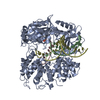
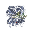
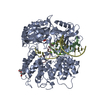
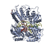
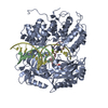
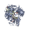
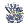
- PDB-6p0c: Human DNA Ligase 1 Bound to an Adenylated, hydroxyl terminated DN... -
+ Open data

- Basic information
| Entry | Database: PDB / ID: 6p0c | ||||||
|---|---|---|---|---|---|---|---|
| Title | Human DNA Ligase 1 Bound to an Adenylated, hydroxyl terminated DNA nick in EDTA | ||||||
 Components Components |
| ||||||
 Keywords Keywords | LIGASE/DNA /  protein-DNA complex / phosphotransferase / OB fold / DNA Binding domain / Adenylation Domain / metalloenzyme / LIGASE / LIGASE-DNA complex protein-DNA complex / phosphotransferase / OB fold / DNA Binding domain / Adenylation Domain / metalloenzyme / LIGASE / LIGASE-DNA complex | ||||||
| Function / homology |  Function and homology information Function and homology informationOkazaki fragment processing involved in mitotic DNA replication / DNA ligase activity / DNA ligase (ATP) / DNA ligase (ATP) activity / Processive synthesis on the lagging strand / DNA ligation / Processive synthesis on the C-strand of the telomere / Mismatch repair (MMR) directed by MSH2:MSH3 (MutSbeta) / Mismatch repair (MMR) directed by MSH2:MSH6 (MutSalpha) / lagging strand elongation ...Okazaki fragment processing involved in mitotic DNA replication / DNA ligase activity / DNA ligase (ATP) / DNA ligase (ATP) activity / Processive synthesis on the lagging strand / DNA ligation / Processive synthesis on the C-strand of the telomere / Mismatch repair (MMR) directed by MSH2:MSH3 (MutSbeta) / Mismatch repair (MMR) directed by MSH2:MSH6 (MutSalpha) / lagging strand elongation / DNA biosynthetic process / Early Phase of HIV Life Cycle / PCNA-Dependent Long Patch Base Excision Repair / POLB-Dependent Long Patch Base Excision Repair / mismatch repair / anatomical structure morphogenesis / base-excision repair, gap-filling / Gap-filling DNA repair synthesis and ligation in GG-NER / base-excision repair / Gap-filling DNA repair synthesis and ligation in TC-NER / DNA recombination / cell division / intracellular membrane-bounded organelle / DNA repair / mitochondrion / DNA binding / nucleoplasm / ATP binding / metal ion binding / nucleusSimilarity search - Function | ||||||
| Biological species |  Homo sapiens (human) Homo sapiens (human)synthetic construct (others) | ||||||
| Method | X-RAY DIFFRACTION / SYNCHROTRON / MOLECULAR REPLACEMENT / Resolution: 1.55 Å | ||||||
 Authors Authors | Schellenberg, M.J. / Tumbale, P.S. / Riccio, A.A. / Williams, R.S. | ||||||
| Funding support |  United States, 1items United States, 1items
| ||||||
 Citation Citation | Journal: Nat Commun / Year: 2019 Title: Two-tiered enforcement of high-fidelity DNA ligation. Authors: Tumbale, P.P. / Jurkiw, T.J. / Schellenberg, M.J. / Riccio, A.A. / O'Brien, P.J. / Williams, R.S. | ||||||
| History |
|
- Structure visualization
| Structure viewer | Molecule: MolmilJmol/JSmol |
|---|
- Downloads & links
-Download
| PDBx/mmCIF format | 6p0c.cif.gz | 491.2 KB | Display | PDBx/mmCIF format |
|---|---|---|---|---|
| PDB format | pdb6p0c.ent.gz | 397.4 KB | Display | PDB format |
| PDBx/mmJSON format | 6p0c.json.gz | Tree view | PDBx/mmJSON format | |
| Others |  Other downloads Other downloads |
-Validation report
| Arichive directory | https://data.pdbj.org/pub/pdb/validation_reports/p0/6p0cftp://data.pdbj.org/pub/pdb/validation_reports/p0/6p0c | HTTPS FTP |
|---|
-Related structure data
| Related structure data |  6p09C  6p0aC 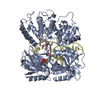 6p0bC 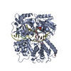 6p0dC  6p0eC  6q1vC 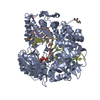 1x9nS S: Starting model for refinement C: citing same article ( |
|---|---|
| Similar structure data |


-Links
 PDBj
PDBj


- Assembly
| Deposited unit | 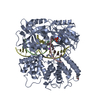
| ||||||||
|---|---|---|---|---|---|---|---|---|---|
| 1 |
| ||||||||
| Unit cell |
|
-Components
-Protein , 1 types, 1 molecules A
| #1: Protein | / DNA ligase I / Polydeoxyribonucleotide synthase [ATP] 1 Mass: 71786.016 Da / Num. of mol.: 1 / Fragment: residues 262-904 Source method: isolated from a genetically manipulated source Source: (gene. exp.) Homo sapiens (human) / Gene: LIG1 / Plasmid: pMCSG7 / Production host:  Escherichia coli (E. coli) / References: UniProt: P18858, DNA ligase (ATP) Escherichia coli (E. coli) / References: UniProt: P18858, DNA ligase (ATP) |
|---|
-DNA chain , 3 types, 3 molecules BCD
| #2: DNA chain | Mass: 3365.196 Da / Num. of mol.: 1 / Source method: obtained synthetically / Source: (synth.) synthetic construct (others) |
|---|---|
| #3: DNA chain | Mass: 2138.423 Da / Num. of mol.: 1 / Source method: obtained synthetically / Source: (synth.) synthetic construct (others) |
| #4: DNA chain | Mass: 5486.557 Da / Num. of mol.: 1 / Source method: obtained synthetically / Source: (synth.) synthetic construct (others) |
-Non-polymers , 4 types, 1117 molecules 


| #5: Chemical | ChemComp-PEG / Diethylene glycol Mass: 106.120 Da / Num. of mol.: 1 / Source method: obtained synthetically / Formula: C4H10O3 Mass: 106.120 Da / Num. of mol.: 1 / Source method: obtained synthetically / Formula: C4H10O3 |
|---|---|
| #6: Chemical | ChemComp-AMP / Adenosine monophosphate Mass: 347.221 Da / Num. of mol.: 1 / Source method: obtained synthetically / Formula: C10H14N5O7P / Comment: AMP*YM Mass: 347.221 Da / Num. of mol.: 1 / Source method: obtained synthetically / Formula: C10H14N5O7P / Comment: AMP*YM |
| #7: Chemical | ChemComp-NA /  Mass: 22.990 Da / Num. of mol.: 1 / Source method: obtained synthetically / Formula: Na Mass: 22.990 Da / Num. of mol.: 1 / Source method: obtained synthetically / Formula: Na |
| #8: Water | ChemComp-HOH / WaterMass: 18.015 Da / Num. of mol.: 1114 / Source method: isolated from a natural source / Formula: H2O |
-Experimental details
-Experiment
| Experiment | Method: X-RAY DIFFRACTION / Number of used crystals: 1 |
|---|
- Sample preparation
| Crystal | Density Matthews: 2.53 Å3/Da / Density % sol: 51.29 % |
|---|---|
| Crystal grow | Temperature: 293 K / Method: vapor diffusion, sitting drop / pH: 6 Details: 100 mM MES, 100 mM Lithium Acetate, 15% (w/v) PEG 3350 |
-Data collection
| Diffraction | Mean temperature: 100 K / Serial crystal experiment: N | |||||||||||||||||||||||||||||||||||||||||||||||||||||||||||||||||||||||||||||||||||||||||||||||||||
|---|---|---|---|---|---|---|---|---|---|---|---|---|---|---|---|---|---|---|---|---|---|---|---|---|---|---|---|---|---|---|---|---|---|---|---|---|---|---|---|---|---|---|---|---|---|---|---|---|---|---|---|---|---|---|---|---|---|---|---|---|---|---|---|---|---|---|---|---|---|---|---|---|---|---|---|---|---|---|---|---|---|---|---|---|---|---|---|---|---|---|---|---|---|---|---|---|---|---|---|---|
| Diffraction source | Source: SYNCHROTRON / Site: APS / Beamline: 22-ID / Wavelength: 1 Å | |||||||||||||||||||||||||||||||||||||||||||||||||||||||||||||||||||||||||||||||||||||||||||||||||||
| Detector | Type: DECTRIS EIGER X 16M / Detector: PIXEL / Date: Dec 15, 2015 | |||||||||||||||||||||||||||||||||||||||||||||||||||||||||||||||||||||||||||||||||||||||||||||||||||
| Radiation | Protocol: SINGLE WAVELENGTH / Monochromatic (M) / Laue (L): M / Scattering type: x-ray | |||||||||||||||||||||||||||||||||||||||||||||||||||||||||||||||||||||||||||||||||||||||||||||||||||
| Radiation wavelength | Wavelength: 1 Å / Relative weight: 1 | |||||||||||||||||||||||||||||||||||||||||||||||||||||||||||||||||||||||||||||||||||||||||||||||||||
| Reflection | Resolution: 1.55→50 Å / Num. obs: 120809 / % possible obs: 97.9 % / Redundancy: 6.9 % / Biso Wilson estimate: 19.56 Å2 / Rmerge(I) obs: 0.11 / Rpim(I) all: 0.045 / Rrim(I) all: 0.119 / Χ2: 1.01 / Net I/σ(I): 8.6 / Num. measured all: 831310 | |||||||||||||||||||||||||||||||||||||||||||||||||||||||||||||||||||||||||||||||||||||||||||||||||||
| Reflection shell | Diffraction-ID: 1
|
- Processing
| Software |
| |||||||||||||||||||||||||||||||||||||||||||||||||||||||||||||||||||||||||||||||||||||||||||||||||||||||||||||||||||||||||||||||||||||||||||||||||||||||||||||||||||||||||||||||||||||||||||||||||||||||||||||||||||||||||
|---|---|---|---|---|---|---|---|---|---|---|---|---|---|---|---|---|---|---|---|---|---|---|---|---|---|---|---|---|---|---|---|---|---|---|---|---|---|---|---|---|---|---|---|---|---|---|---|---|---|---|---|---|---|---|---|---|---|---|---|---|---|---|---|---|---|---|---|---|---|---|---|---|---|---|---|---|---|---|---|---|---|---|---|---|---|---|---|---|---|---|---|---|---|---|---|---|---|---|---|---|---|---|---|---|---|---|---|---|---|---|---|---|---|---|---|---|---|---|---|---|---|---|---|---|---|---|---|---|---|---|---|---|---|---|---|---|---|---|---|---|---|---|---|---|---|---|---|---|---|---|---|---|---|---|---|---|---|---|---|---|---|---|---|---|---|---|---|---|---|---|---|---|---|---|---|---|---|---|---|---|---|---|---|---|---|---|---|---|---|---|---|---|---|---|---|---|---|---|---|---|---|---|---|---|---|---|---|---|---|---|---|---|---|---|---|---|---|---|
| Refinement | Method to determine structure: MOLECULAR REPLACEMENT Starting model: 1X9N Resolution: 1.55→33.602 Å / SU ML: 0.16 / Cross valid method: THROUGHOUT / σ(F): 1.33 / Phase error: 18.33
| |||||||||||||||||||||||||||||||||||||||||||||||||||||||||||||||||||||||||||||||||||||||||||||||||||||||||||||||||||||||||||||||||||||||||||||||||||||||||||||||||||||||||||||||||||||||||||||||||||||||||||||||||||||||||
| Solvent computation | Shrinkage radii: 0.9 Å / VDW probe radii: 1.11 Å | |||||||||||||||||||||||||||||||||||||||||||||||||||||||||||||||||||||||||||||||||||||||||||||||||||||||||||||||||||||||||||||||||||||||||||||||||||||||||||||||||||||||||||||||||||||||||||||||||||||||||||||||||||||||||
| Displacement parameters | Biso max: 117 Å2 / Biso mean: 30.8197 Å2 / Biso min: 12.15 Å2 | |||||||||||||||||||||||||||||||||||||||||||||||||||||||||||||||||||||||||||||||||||||||||||||||||||||||||||||||||||||||||||||||||||||||||||||||||||||||||||||||||||||||||||||||||||||||||||||||||||||||||||||||||||||||||
| Refinement step | Cycle: final / Resolution: 1.55→33.602 Å
| |||||||||||||||||||||||||||||||||||||||||||||||||||||||||||||||||||||||||||||||||||||||||||||||||||||||||||||||||||||||||||||||||||||||||||||||||||||||||||||||||||||||||||||||||||||||||||||||||||||||||||||||||||||||||
| Refine LS restraints |
| |||||||||||||||||||||||||||||||||||||||||||||||||||||||||||||||||||||||||||||||||||||||||||||||||||||||||||||||||||||||||||||||||||||||||||||||||||||||||||||||||||||||||||||||||||||||||||||||||||||||||||||||||||||||||
| LS refinement shell | Refine-ID: X-RAY DIFFRACTION / Rfactor Rfree error: 0 / Total num. of bins used: 30
|
