 Movie
Movie Controller


+ Open data

- Basic information
| Entry | Database: PDB / ID: 7kqx | ||||||
|---|---|---|---|---|---|---|---|
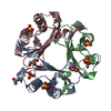
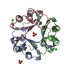
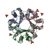
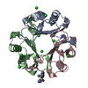
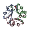
| Title | MIF Y99C homotrimeric mutant | ||||||
 Components Components | Macrophage migration inhibitory factor | ||||||
 Keywords Keywords | ISOMERASE / MIF / mutant | ||||||
| Function / homology |  Function and homology information Function and homology informationpositive regulation of prostaglandin secretion involved in immune response / positive regulation of myeloid leukocyte cytokine production involved in immune response / phenylpyruvate tautomerase / L-dopachrome isomerase / dopachrome isomerase activity / phenylpyruvate tautomerase activity / positive regulation of lipopolysaccharide-mediated signaling pathway / cytokine receptor binding / positive regulation of arachidonic acid secretion / negative regulation of myeloid cell apoptotic process ...positive regulation of prostaglandin secretion involved in immune response / positive regulation of myeloid leukocyte cytokine production involved in immune response / phenylpyruvate tautomerase / L-dopachrome isomerase / dopachrome isomerase activity / phenylpyruvate tautomerase activity / positive regulation of lipopolysaccharide-mediated signaling pathway / cytokine receptor binding / positive regulation of arachidonic acid secretion / negative regulation of myeloid cell apoptotic process / negative regulation of mature B cell apoptotic process / negative regulation of macrophage chemotaxis / positive regulation of chemokine (C-X-C motif) ligand 2 production / carboxylic acid metabolic process / prostaglandin biosynthetic process / negative regulation of protein metabolic process / regulation of macrophage activation / negative regulation of intrinsic apoptotic signaling pathway in response to DNA damage by p53 class mediator / chemoattractant activity / positive regulation of protein kinase A signaling / protein homotrimerization / negative regulation of cellular senescence / negative regulation of DNA damage response, signal transduction by p53 class mediator / DNA damage response, signal transduction by p53 class mediator / positive regulation of B cell proliferation / positive regulation of phosphorylation / Gene and protein expression by JAK-STAT signaling after Interleukin-12 stimulation / negative regulation of cell migration / positive regulation of cytokine production / cytokine activity / Cell surface interactions at the vascular wall / positive regulation of tumor necrosis factor production / cellular senescence / positive regulation of peptidyl-tyrosine phosphorylation / positive regulation of fibroblast proliferation / positive regulation of peptidyl-serine phosphorylation / secretory granule lumen / protease binding / vesicle / ficolin-1-rich granule lumen / cell surface receptor signaling pathway / positive regulation of ERK1 and ERK2 cascade / inflammatory response / negative regulation of gene expression / innate immune response / Neutrophil degranulation / positive regulation of cell population proliferation / negative regulation of apoptotic process / cell surface / extracellular space / extracellular exosome / extracellular region / nucleoplasm / identical protein binding / plasma membrane / cytosol / cytoplasmSimilarity search - Function | ||||||
| Biological species |  Homo sapiens (human) Homo sapiens (human) | ||||||
| Method | X-RAY DIFFRACTION / MOLECULAR REPLACEMENT / molecular replacement / Resolution: 1.6 Å | ||||||
 Authors Authors | Manjula, R. / Georgios, P. / Lolis, E.J. | ||||||
| Funding support | 1items
| ||||||
 Citation Citation | Journal: Front Mol Biosci / Year: 2022 Title: A Cysteine Variant at an Allosteric Site Alters MIF Dynamics and Biological Function in Homo- and Heterotrimeric Assemblies. Authors: Skeens, E. / Pantouris, G. / Shah, D. / Manjula, R. / Ombrello, M.J. / Maluf, N.K. / Bhandari, V. / Lisi, G.P. / Lolis, E.J. | ||||||
| History |
|
- Structure visualization
| Structure viewer | Molecule: MolmilJmol/JSmol |
|---|
- Downloads & links
-Download
| PDBx/mmCIF format | 7kqx.cif.gz | 90 KB | Display | PDBx/mmCIF format |
|---|---|---|---|---|
| PDB format | pdb7kqx.ent.gz | 66.8 KB | Display | PDB format |
| PDBx/mmJSON format | 7kqx.json.gz | Tree view | PDBx/mmJSON format | |
| Others |  Other downloads Other downloads |
-Validation report
| Arichive directory | https://data.pdbj.org/pub/pdb/validation_reports/kq/7kqxftp://data.pdbj.org/pub/pdb/validation_reports/kq/7kqx | HTTPS FTP |
|---|
-Related structure data
| Related structure data | 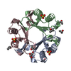 5bs9S S: Starting model for refinement |
|---|---|
| Similar structure data |


-Links
 PDBj
PDBj


- Assembly
| Deposited unit | 
| ||||||||
|---|---|---|---|---|---|---|---|---|---|
| 1 |
| ||||||||
| Unit cell |
|
-Components
| #1: Protein | / MIF / Glycosylation-inhibiting factor / GIF / L-dopachrome isomerase / L-dopachrome tautomerase / ...MIF / Glycosylation-inhibiting factor / GIF / L-dopachrome isomerase / L-dopachrome tautomerase / Phenylpyruvate tautomerase Mass: 12295.024 Da / Num. of mol.: 3 / Mutation: Y99C Source method: isolated from a genetically manipulated source Source: (gene. exp.) Homo sapiens (human) / Gene: MIF, GLIF, MMIF / Production host:  Escherichia coli (E. coli) Escherichia coli (E. coli)References: UniProt: P14174, phenylpyruvate tautomerase, L-dopachrome isomerase#2: Chemical | Sulfate  Mass: 96.063 Da / Num. of mol.: 3 / Source method: obtained synthetically / Formula: SO4 / Feature type: SUBJECT OF INVESTIGATION Mass: 96.063 Da / Num. of mol.: 3 / Source method: obtained synthetically / Formula: SO4 / Feature type: SUBJECT OF INVESTIGATION#3: Chemical | ChemComp-GOL / Glycerol  Mass: 92.094 Da / Num. of mol.: 15 / Source method: obtained synthetically / Formula: C3H8O3 / Feature type: SUBJECT OF INVESTIGATION Mass: 92.094 Da / Num. of mol.: 15 / Source method: obtained synthetically / Formula: C3H8O3 / Feature type: SUBJECT OF INVESTIGATION#4: Chemical | ChemComp-IPA / Isopropyl alcohol  Mass: 60.095 Da / Num. of mol.: 5 / Source method: obtained synthetically / Formula: C3H8O / Feature type: SUBJECT OF INVESTIGATION / Comment: alkaloid*YM Mass: 60.095 Da / Num. of mol.: 5 / Source method: obtained synthetically / Formula: C3H8O / Feature type: SUBJECT OF INVESTIGATION / Comment: alkaloid*YM#5: Water | ChemComp-HOH / | Water Mass: 18.015 Da / Num. of mol.: 365 / Source method: isolated from a natural source / Formula: H2O Mass: 18.015 Da / Num. of mol.: 365 / Source method: isolated from a natural source / Formula: H2OHas ligand of interest | Y | |
|---|
-Experimental details
-Experiment
| Experiment | Method: X-RAY DIFFRACTION / Number of used crystals: 1 |
|---|
- Sample preparation
| Crystal | Density Matthews: 2.72 Å3/Da / Density % sol: 54.78 % |
|---|---|
| Crystal grow | Temperature: 293 K / Method: vapor diffusion, hanging drop / pH: 7.5 Details: 2 M ammonium sulfate, 3% 2-propanol, 0.1 M Tris-HCl, pH 7.5 |
-Data collection
| Diffraction | Mean temperature: 93 K / Serial crystal experiment: N | |||||||||||||||||||||||||||||||||||||||||||||||||||||||||||||||||||||||||||||||||||||||||||||||||||||||||||||||||||||||||||||||||||||||||||||||||||||||||||||||||||||||||||||||||||||||||||||
|---|---|---|---|---|---|---|---|---|---|---|---|---|---|---|---|---|---|---|---|---|---|---|---|---|---|---|---|---|---|---|---|---|---|---|---|---|---|---|---|---|---|---|---|---|---|---|---|---|---|---|---|---|---|---|---|---|---|---|---|---|---|---|---|---|---|---|---|---|---|---|---|---|---|---|---|---|---|---|---|---|---|---|---|---|---|---|---|---|---|---|---|---|---|---|---|---|---|---|---|---|---|---|---|---|---|---|---|---|---|---|---|---|---|---|---|---|---|---|---|---|---|---|---|---|---|---|---|---|---|---|---|---|---|---|---|---|---|---|---|---|---|---|---|---|---|---|---|---|---|---|---|---|---|---|---|---|---|---|---|---|---|---|---|---|---|---|---|---|---|---|---|---|---|---|---|---|---|---|---|---|---|---|---|---|---|---|---|---|---|---|
| Diffraction source | Source: ROTATING ANODE / Type: RIGAKU / Wavelength: 1.5428 Å | |||||||||||||||||||||||||||||||||||||||||||||||||||||||||||||||||||||||||||||||||||||||||||||||||||||||||||||||||||||||||||||||||||||||||||||||||||||||||||||||||||||||||||||||||||||||||||||
| Detector | Type: DECTRIS PILATUS 200K / Detector: PIXEL / Date: Apr 29, 2015 | |||||||||||||||||||||||||||||||||||||||||||||||||||||||||||||||||||||||||||||||||||||||||||||||||||||||||||||||||||||||||||||||||||||||||||||||||||||||||||||||||||||||||||||||||||||||||||||
| Radiation | Protocol: SINGLE WAVELENGTH / Monochromatic (M) / Laue (L): M / Scattering type: x-ray | |||||||||||||||||||||||||||||||||||||||||||||||||||||||||||||||||||||||||||||||||||||||||||||||||||||||||||||||||||||||||||||||||||||||||||||||||||||||||||||||||||||||||||||||||||||||||||||
| Radiation wavelength | Wavelength: 1.5428 Å / Relative weight: 1 | |||||||||||||||||||||||||||||||||||||||||||||||||||||||||||||||||||||||||||||||||||||||||||||||||||||||||||||||||||||||||||||||||||||||||||||||||||||||||||||||||||||||||||||||||||||||||||||
| Reflection | Resolution: 1.6→50 Å / Num. obs: 53669 / % possible obs: 99.6 % / Redundancy: 5.7 % / Biso Wilson estimate: 14.3 Å2 / Rmerge(I) obs: 0.046 / Rpim(I) all: 0.018 / Rrim(I) all: 0.05 / Χ2: 0.93 / Net I/σ(I): 16.3 / Num. measured all: 303731 | |||||||||||||||||||||||||||||||||||||||||||||||||||||||||||||||||||||||||||||||||||||||||||||||||||||||||||||||||||||||||||||||||||||||||||||||||||||||||||||||||||||||||||||||||||||||||||||
| Reflection shell | Diffraction-ID: 1
|
-Phasing
| Phasing | Method: molecular replacement | ||||||
|---|---|---|---|---|---|---|---|
| Phasing MR |
|
- Processing
| Software |
| |||||||||||||||||||||||||||||||||||||||||||||||||||||||||||||||||||||||||||||||||||||||||||||||||||||||||||||||||||||||||||||||||||||
|---|---|---|---|---|---|---|---|---|---|---|---|---|---|---|---|---|---|---|---|---|---|---|---|---|---|---|---|---|---|---|---|---|---|---|---|---|---|---|---|---|---|---|---|---|---|---|---|---|---|---|---|---|---|---|---|---|---|---|---|---|---|---|---|---|---|---|---|---|---|---|---|---|---|---|---|---|---|---|---|---|---|---|---|---|---|---|---|---|---|---|---|---|---|---|---|---|---|---|---|---|---|---|---|---|---|---|---|---|---|---|---|---|---|---|---|---|---|---|---|---|---|---|---|---|---|---|---|---|---|---|---|---|---|---|
| Refinement | Method to determine structure: MOLECULAR REPLACEMENT Starting model: 5BS9 Resolution: 1.6→48.1 Å / SU ML: 0.14 / Cross valid method: FREE R-VALUE / σ(F): 1.34 / Phase error: 17.32 / Stereochemistry target values: ML
| |||||||||||||||||||||||||||||||||||||||||||||||||||||||||||||||||||||||||||||||||||||||||||||||||||||||||||||||||||||||||||||||||||||
| Solvent computation | Shrinkage radii: 0.9 Å / VDW probe radii: 1.11 Å / Solvent model: FLAT BULK SOLVENT MODEL | |||||||||||||||||||||||||||||||||||||||||||||||||||||||||||||||||||||||||||||||||||||||||||||||||||||||||||||||||||||||||||||||||||||
| Refinement step | Cycle: LAST / Resolution: 1.6→48.1 Å
| |||||||||||||||||||||||||||||||||||||||||||||||||||||||||||||||||||||||||||||||||||||||||||||||||||||||||||||||||||||||||||||||||||||
| Refine LS restraints |
| |||||||||||||||||||||||||||||||||||||||||||||||||||||||||||||||||||||||||||||||||||||||||||||||||||||||||||||||||||||||||||||||||||||
| LS refinement shell |
|
