 Movie
Movie Controller


+ Open data

- Basic information
| Entry | Database: PDB / ID: 7kb6 | ||||||
|---|---|---|---|---|---|---|---|
| Title | Co-crystal structure of alpha glucosidase with compound 7 | ||||||
 Components Components |
| ||||||
 Keywords Keywords |  HYDROLASE / Alpha glucosidase II / Endoplasmic reticulum / Inhibitor complex HYDROLASE / Alpha glucosidase II / Endoplasmic reticulum / Inhibitor complex | ||||||
| Function / homology |  Function and homology information Function and homology informationmannosyl-oligosaccharide alpha-1,3-glucosidase / glucosidase II complex / Regulation of Insulin-like Growth Factor (IGF) transport and uptake by Insulin-like Growth Factor Binding Proteins (IGFBPs) / Post-translational protein phosphorylation / N-glycan processing / liver development / negative regulation of neuron projection development / in utero embryonic development / intracellular membrane-bounded organelle / calcium ion binding ...mannosyl-oligosaccharide alpha-1,3-glucosidase / glucosidase II complex / Regulation of Insulin-like Growth Factor (IGF) transport and uptake by Insulin-like Growth Factor Binding Proteins (IGFBPs) / Post-translational protein phosphorylation / N-glycan processing / liver development / negative regulation of neuron projection development / in utero embryonic development / intracellular membrane-bounded organelle / calcium ion binding / protein-containing complex binding / endoplasmic reticulum / RNA bindingSimilarity search - Function | ||||||
| Biological species |  Mus musculus (house mouse) Mus musculus (house mouse) | ||||||
| Method | X-RAY DIFFRACTION / SYNCHROTRON / MOLECULAR REPLACEMENT / Resolution: 2.2 Å | ||||||
 Authors Authors | Karade, S.S. / Mariuzza, R.A. | ||||||
 Citation Citation | Journal: J.Med.Chem. / Year: 2021 Title: N-Substituted Valiolamine Derivatives as Potent Inhibitors of Endoplasmic Reticulum alpha-Glucosidases I and II with Antiviral Activity. Authors: Karade, S.S. / Hill, M.L. / Kiappes, J.L. / Manne, R. / Aakula, B. / Zitzmann, N. / Warfield, K.L. / Treston, A.M. / Mariuzza, R.A. | ||||||
| History |
|
- Structure visualization
| Structure viewer | Molecule: MolmilJmol/JSmol |
|---|
- Downloads & links
-Download
| PDBx/mmCIF format | 7kb6.cif.gz | 539.9 KB | Display | PDBx/mmCIF format |
|---|---|---|---|---|
| PDB format | pdb7kb6.ent.gz | 339.4 KB | Display | PDB format |
| PDBx/mmJSON format | 7kb6.json.gz | Tree view | PDBx/mmJSON format | |
| Others |  Other downloads Other downloads |
-Validation report
| Arichive directory | https://data.pdbj.org/pub/pdb/validation_reports/kb/7kb6ftp://data.pdbj.org/pub/pdb/validation_reports/kb/7kb6 | HTTPS FTP |
|---|
-Related structure data
| Related structure data | 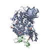 7jtyC  7k9nC 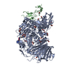 7k9oC 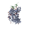 7k9qC  7k9tC 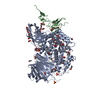 7kadC  7kb8C  7kbjC  7kbrC  7kryC  7l9eC  5f0eS S: Starting model for refinement C: citing same article ( |
|---|---|
| Similar structure data |


-Links
 PDBj
PDBj


- Assembly
| Deposited unit | 
| ||||||||||||
|---|---|---|---|---|---|---|---|---|---|---|---|---|---|
| 1 | 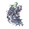
| ||||||||||||
| 2 | 
| ||||||||||||
| Unit cell |
|
-Components
-Protein , 2 types, 4 molecules ACBD
| #1: Protein | Mass: 110654.062 Da / Num. of mol.: 2 / Mutation: N97D Source method: isolated from a genetically manipulated source Source: (gene. exp.) Mus musculus (house mouse) / Gene: Ganab, G2an, Kiaa0088 / Cell line (production host): Expi-HEK293 / Production host:  Homo sapiens (human) Homo sapiens (human)References: UniProt: Q8BHN3-2, mannosyl-oligosaccharide alpha-1,3-glucosidase #2: Protein | Glucosidases / 80K-H protein / Glucosidase II subunit beta / Protein kinase C substrate 60.1 kDa protein heavy chain / PKCSHMass: 62307.430 Da / Num. of mol.: 2 Source method: isolated from a genetically manipulated source Source: (gene. exp.) Mus musculus (house mouse) / Gene: Prkcsh / Cell line (production host): Expi-HEK293 / Production host: Homo sapiens (human) / References: UniProt: O08795 |
|---|
-Non-polymers , 8 types, 963 molecules 


| #3: Chemical | ChemComp-PGE / Polyethylene glycol Mass: 150.173 Da / Num. of mol.: 9 / Source method: obtained synthetically / Formula: C6H14O4 Mass: 150.173 Da / Num. of mol.: 9 / Source method: obtained synthetically / Formula: C6H14O4#4: Chemical | ChemComp-EDO / Ethylene glycol Mass: 62.068 Da / Num. of mol.: 45 / Source method: obtained synthetically / Formula: C2H6O2 Mass: 62.068 Da / Num. of mol.: 45 / Source method: obtained synthetically / Formula: C2H6O2#5: Chemical | ChemComp-PEG / Diethylene glycol Mass: 106.120 Da / Num. of mol.: 14 / Source method: obtained synthetically / Formula: C4H10O3 Mass: 106.120 Da / Num. of mol.: 14 / Source method: obtained synthetically / Formula: C4H10O3#6: Chemical |  Mass: 491.537 Da / Num. of mol.: 2 / Source method: obtained synthetically / Formula: C23H33N5O7 / Feature type: SUBJECT OF INVESTIGATION Mass: 491.537 Da / Num. of mol.: 2 / Source method: obtained synthetically / Formula: C23H33N5O7 / Feature type: SUBJECT OF INVESTIGATION#7: Chemical | ChemComp-SO4 / Sulfate Mass: 96.063 Da / Num. of mol.: 10 / Source method: obtained synthetically / Formula: SO4 Mass: 96.063 Da / Num. of mol.: 10 / Source method: obtained synthetically / Formula: SO4#8: Chemical | ChemComp-CA /  Mass: 40.078 Da / Num. of mol.: 4 / Source method: obtained synthetically / Formula: Ca Mass: 40.078 Da / Num. of mol.: 4 / Source method: obtained synthetically / Formula: Ca#9: Chemical | Polyethylene glycol Mass: 194.226 Da / Num. of mol.: 2 / Source method: obtained synthetically / Formula: C8H18O5 / Comment: precipitant*YM Mass: 194.226 Da / Num. of mol.: 2 / Source method: obtained synthetically / Formula: C8H18O5 / Comment: precipitant*YM#10: Water | ChemComp-HOH / | WaterMass: 18.015 Da / Num. of mol.: 877 / Source method: isolated from a natural source / Formula: H2O |
|---|
-Details
| Has ligand of interest | Y |
|---|
-Experimental details
-Experiment
| Experiment | Method: X-RAY DIFFRACTION / Number of used crystals: 1 |
|---|
- Sample preparation
| Crystal | Density Matthews: 2.12 Å3/Da / Density % sol: 41.97 % |
|---|---|
| Crystal grow | Temperature: 297 K / Method: vapor diffusion, sitting drop / pH: 7 Details: 0.09M NPS, 0.1M Buffer system 1 pH 7.0, 29.0%v/v P500MME_P20K (Morpheus screen, condition C1) |
-Data collection
| Diffraction | Mean temperature: 100 K / Serial crystal experiment: N |
|---|---|
| Diffraction source | Source: SYNCHROTRON / Site: APS  / Beamline: 23-ID-B / Wavelength: 0.9793 Å / Beamline: 23-ID-B / Wavelength: 0.9793 Å |
| Detector | Type: DECTRIS EIGER X 16M / Detector: PIXEL / Date: Apr 8, 2020 / Details: Adjustable focus K-B pair Si plus Pt, Rh coatings |
| Radiation | Monochromator: Double crystal cryo-cooled Si(111) / Protocol: SINGLE WAVELENGTH / Monochromatic (M) / Laue (L): M / Scattering type: x-ray |
| Radiation wavelength | Wavelength: 0.9793 Å / Relative weight: 1 |
| Reflection | Resolution: 2.2→44.56 Å / Num. obs: 140562 / % possible obs: 97.47 % / Redundancy: 6.4 % / Biso Wilson estimate: 38.09 Å2 / CC1/2: 0.993 / CC star: 0.998 / Rmerge(I) obs: 0.1286 / Rpim(I) all: 0.0524 / Rrim(I) all: 0.1392 / Net I/σ(I): 9.2 |
| Reflection shell | Resolution: 2.2→2.279 Å / Rmerge(I) obs: 0.9757 / Mean I/σ(I) obs: 1.13 / Num. unique obs: 13540 / CC1/2: 0.654 / CC star: 0.889 / Rpim(I) all: 0.4064 / Rrim(I) all: 1.06 / % possible all: 93.75 |
- Processing
| Software |
| |||||||||||||||||||||||||||||||||||||||||||||||||||||||||||||||||||||||||||||||||||||||||||||||||||||||||
|---|---|---|---|---|---|---|---|---|---|---|---|---|---|---|---|---|---|---|---|---|---|---|---|---|---|---|---|---|---|---|---|---|---|---|---|---|---|---|---|---|---|---|---|---|---|---|---|---|---|---|---|---|---|---|---|---|---|---|---|---|---|---|---|---|---|---|---|---|---|---|---|---|---|---|---|---|---|---|---|---|---|---|---|---|---|---|---|---|---|---|---|---|---|---|---|---|---|---|---|---|---|---|---|---|---|---|
| Refinement | Method to determine structure: MOLECULAR REPLACEMENT Starting model: 5F0E Resolution: 2.2→44.56 Å / SU ML: 0.2552 / Cross valid method: FREE R-VALUE / σ(F): 1.96 / Phase error: 21.7639 Stereochemistry target values: GeoStd + Monomer Library + CDL v1.2
| |||||||||||||||||||||||||||||||||||||||||||||||||||||||||||||||||||||||||||||||||||||||||||||||||||||||||
| Solvent computation | Shrinkage radii: 0.9 Å / VDW probe radii: 1.11 Å / Solvent model: FLAT BULK SOLVENT MODEL | |||||||||||||||||||||||||||||||||||||||||||||||||||||||||||||||||||||||||||||||||||||||||||||||||||||||||
| Displacement parameters | Biso mean: 43.77 Å2 | |||||||||||||||||||||||||||||||||||||||||||||||||||||||||||||||||||||||||||||||||||||||||||||||||||||||||
| Refinement step | Cycle: LAST / Resolution: 2.2→44.56 Å
| |||||||||||||||||||||||||||||||||||||||||||||||||||||||||||||||||||||||||||||||||||||||||||||||||||||||||
| Refine LS restraints |
| |||||||||||||||||||||||||||||||||||||||||||||||||||||||||||||||||||||||||||||||||||||||||||||||||||||||||
| LS refinement shell |
|
