 Movie
Movie Controller

[English] 日本語
 Yorodumi
- PDB-7k5n: Ligand binding domain (tandem PAS/dCache) of Aeromonas caviae dig... -
+ Open data

- Basic information
| Entry | Database: PDB / ID: 7k5n | ||||||||||||
|---|---|---|---|---|---|---|---|---|---|---|---|---|---|
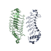
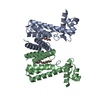
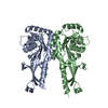
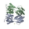
| Title | Ligand binding domain (tandem PAS/dCache) of Aeromonas caviae diguanylate cyclase with proline bound | ||||||||||||
 Components Components | Sensor domain-containing diguanylate cyclase | ||||||||||||
 Keywords Keywords |  SIGNALING PROTEIN / PAS / Cache / bacterial inner membrane bound / amino acid binder SIGNALING PROTEIN / PAS / Cache / bacterial inner membrane bound / amino acid binder | ||||||||||||
| Function / homology |  Function and homology information Function and homology information | ||||||||||||
| Biological species |  Aeromonas caviae (bacteria) Aeromonas caviae (bacteria) | ||||||||||||
| Method | X-RAY DIFFRACTION / SYNCHROTRON / MOLECULAR REPLACEMENT / Resolution: 1.8 Å | ||||||||||||
 Authors Authors | Sweeney, E.G. / Remington, S.J. | ||||||||||||
| Funding support |  United States, 3items United States, 3items
| ||||||||||||
 Citation Citation | Journal: Cell Host Microbe / Year: 2021 Title: Host-emitted amino acid cues regulate bacterial chemokinesis to enhance colonization. Authors: Robinson, C.D. / Sweeney, E.G. / Ngo, J. / Ma, E. / Perkins, A. / Smith, T.J. / Fernandez, N.L. / Waters, C.M. / Remington, S.J. / Bohannan, B.J.M. / Guillemin, K. | ||||||||||||
| History |
|
- Structure visualization
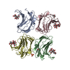
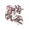
| Structure viewer | Molecule: MolmilJmol/JSmol |
|---|
- Downloads & links
-Download
| PDBx/mmCIF format | 7k5n.cif.gz | 103.2 KB | Display | PDBx/mmCIF format |
|---|---|---|---|---|
| PDB format | pdb7k5n.ent.gz | 78.7 KB | Display | PDB format |
| PDBx/mmJSON format | 7k5n.json.gz | Tree view | PDBx/mmJSON format | |
| Others |  Other downloads Other downloads |
-Validation report
| Arichive directory | https://data.pdbj.org/pub/pdb/validation_reports/k5/7k5nftp://data.pdbj.org/pub/pdb/validation_reports/k5/7k5n | HTTPS FTP |
|---|
-Related structure data
| Related structure data | 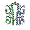 3c8cS S: Starting model for refinement |
|---|---|
| Similar structure data |
-Links
 PDBj
PDBj


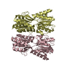
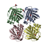
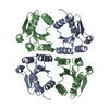
- Assembly
| Deposited unit | 
| ||||||||
|---|---|---|---|---|---|---|---|---|---|
| 1 | 
| ||||||||
| Unit cell |
|
-Components
| #1: Protein | Mass: 31456.561 Da / Num. of mol.: 1 Source method: isolated from a genetically manipulated source Details: zebrafish isolate Guillemin lab, ZOR0002 / Source: (gene. exp.) Aeromonas caviae (bacteria) / Gene: C0708_15830, C1C92_04435 / Plasmid: pBH / Production host: Escherichia coli BL21(DE3) (bacteria) / Strain (production host): BL21(DE3) / References: UniProt: A0A3S5WQC2 |
|---|---|
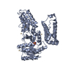
| #2: Chemical | ChemComp-PRO / Proline  Type: L-peptide linking / Mass: 115.130 Da / Num. of mol.: 1 / Source method: obtained synthetically / Formula: C5H9NO2 / Feature type: SUBJECT OF INVESTIGATION Type: L-peptide linking / Mass: 115.130 Da / Num. of mol.: 1 / Source method: obtained synthetically / Formula: C5H9NO2 / Feature type: SUBJECT OF INVESTIGATION |
| #3: Chemical | ChemComp-GOL / Glycerol  Mass: 92.094 Da / Num. of mol.: 1 / Source method: obtained synthetically / Formula: C3H8O3 Mass: 92.094 Da / Num. of mol.: 1 / Source method: obtained synthetically / Formula: C3H8O3 |
| #4: Water | ChemComp-HOH / Water Mass: 18.015 Da / Num. of mol.: 30 / Source method: isolated from a natural source / Formula: H2O Mass: 18.015 Da / Num. of mol.: 30 / Source method: isolated from a natural source / Formula: H2O |
| Has ligand of interest | Y |
-Experimental details
-Experiment
| Experiment | Method: X-RAY DIFFRACTION / Number of used crystals: 1 |
|---|
- Sample preparation
| Crystal | Density Matthews: 2.43 Å3/Da / Density % sol: 44.5 % |
|---|---|
| Crystal grow | Temperature: 293 K / Method: vapor diffusion, hanging drop / pH: 4.5 Details: crystal growth conditions: 80 mM sodium acetate trihydrate pH 4.5 and 1.4 - 1.5 M sodium formate. cryoprotectant was the crystal growth conditions plus 10 mM proline and 20% glycerol. ...Details: crystal growth conditions: 80 mM sodium acetate trihydrate pH 4.5 and 1.4 - 1.5 M sodium formate. cryoprotectant was the crystal growth conditions plus 10 mM proline and 20% glycerol. crystals were briefly (< 1min) swished through cryoprotectant before flash frozen in liquid nitrogen Temp details: room temperature |
-Data collection
| Diffraction | Mean temperature: 80 K / Ambient temp details: collected under cryostream / Serial crystal experiment: N |
|---|---|
| Diffraction source | Source: SYNCHROTRON / Site: ALS / Beamline: 5.0.2 / Wavelength: 1 Å |
| Detector | Type: DECTRIS PILATUS3 S 6M / Detector: PIXEL / Date: Mar 31, 2019 |
| Radiation | Protocol: SINGLE WAVELENGTH / Monochromatic (M) / Laue (L): M / Scattering type: x-ray |
| Radiation wavelength | Wavelength: 1 Å / Relative weight: 1 |
| Reflection | Resolution: 1.8→50 Å / Num. obs: 26946 / % possible obs: 99.99 % / Redundancy: 20 % / CC1/2: 0.99 / CC star: 0.997 / Rmerge(I) obs: 0.063 / Rpim(I) all: 0.064 / Rrim(I) all: 0.089 / Net I/σ(I): 30 |
| Reflection shell | Resolution: 1.8→1.83 Å / Num. unique obs: 1304 / CC1/2: 0.208 / CC star: 0.586 |
- Processing
| Software |
| |||||||||||||||||||||||||||||||||||||||||||||||||||||||||||||||||||||||||||||
|---|---|---|---|---|---|---|---|---|---|---|---|---|---|---|---|---|---|---|---|---|---|---|---|---|---|---|---|---|---|---|---|---|---|---|---|---|---|---|---|---|---|---|---|---|---|---|---|---|---|---|---|---|---|---|---|---|---|---|---|---|---|---|---|---|---|---|---|---|---|---|---|---|---|---|---|---|---|---|
| Refinement | Method to determine structure: MOLECULAR REPLACEMENT Starting model: 3c8c Resolution: 1.8→30.47 Å / SU ML: 0.36 / Cross valid method: THROUGHOUT / σ(F): 1.33 / Phase error: 39.12 / Stereochemistry target values: ML
| |||||||||||||||||||||||||||||||||||||||||||||||||||||||||||||||||||||||||||||
| Solvent computation | Shrinkage radii: 0.9 Å / VDW probe radii: 1.11 Å / Solvent model: FLAT BULK SOLVENT MODEL | |||||||||||||||||||||||||||||||||||||||||||||||||||||||||||||||||||||||||||||
| Displacement parameters | Biso max: 141.91 Å2 / Biso mean: 62.9754 Å2 / Biso min: 35.5 Å2 | |||||||||||||||||||||||||||||||||||||||||||||||||||||||||||||||||||||||||||||
| Refinement step | Cycle: final / Resolution: 1.8→30.47 Å
| |||||||||||||||||||||||||||||||||||||||||||||||||||||||||||||||||||||||||||||
| LS refinement shell | Refine-ID: X-RAY DIFFRACTION / Rfactor Rfree error: 0 / Total num. of bins used: 10
|
