 Movie
Movie Controller


+ Open data

- Basic information
| Entry | Database: PDB / ID: 1jhz | ||||||
|---|---|---|---|---|---|---|---|
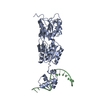
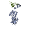
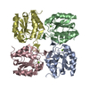
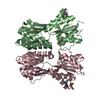
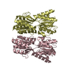
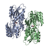
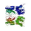
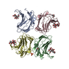
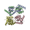
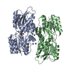
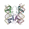
| Title | Purine Repressor Mutant Corepressor Binding Domain Structure | ||||||
 Components Components | PURINE NUCLEOTIDE SYNTHESIS REPRESSOR | ||||||
 Keywords Keywords |  TRANSCRIPTION / corepressor binding domain / purine repressor / allosteric regulation / DNA-binding protein / purine biosynthesis TRANSCRIPTION / corepressor binding domain / purine repressor / allosteric regulation / DNA-binding protein / purine biosynthesis | ||||||
| Function / homology |  Function and homology informationguanine binding / negative regulation of purine nucleotide biosynthetic process / purine nucleotide biosynthetic process / DNA-binding transcription repressor activity / transcription cis-regulatory region binding / DNA-binding transcription factor activity / negative regulation of DNA-templated transcription / regulation of DNA-templated transcription / protein homodimerization activity / cytosol Function and homology informationguanine binding / negative regulation of purine nucleotide biosynthetic process / purine nucleotide biosynthetic process / DNA-binding transcription repressor activity / transcription cis-regulatory region binding / DNA-binding transcription factor activity / negative regulation of DNA-templated transcription / regulation of DNA-templated transcription / protein homodimerization activity / cytosolSimilarity search - Function | ||||||
| Biological species |  Escherichia coli (E. coli) Escherichia coli (E. coli) | ||||||
| Method | X-RAY DIFFRACTION / SYNCHROTRON / MOLECULAR REPLACEMENT / Resolution: 2.4 Å | ||||||
 Authors Authors | Huffman, J.L. / Lu, F. / Zalkin, H. / Brennan, R.G. | ||||||
 Citation Citation | Journal: Biochemistry / Year: 2002 Title: Role of residue 147 in the gene regulatory function of the Escherichia coli purine repressor. Authors: Huffman, J.L. / Lu, F. / Zalkin, H. / Brennan, R.G. | ||||||
| History |
|
- Structure visualization
| Structure viewer | Molecule: MolmilJmol/JSmol |
|---|
- Downloads & links
-Download
| PDBx/mmCIF format | 1jhz.cif.gz | 122.7 KB | Display | PDBx/mmCIF format |
|---|---|---|---|---|
| PDB format | pdb1jhz.ent.gz | 95.1 KB | Display | PDB format |
| PDBx/mmJSON format | 1jhz.json.gz | Tree view | PDBx/mmJSON format | |
| Others |  Other downloads Other downloads |
-Validation report
| Arichive directory | https://data.pdbj.org/pub/pdb/validation_reports/jh/1jhzftp://data.pdbj.org/pub/pdb/validation_reports/jh/1jhz | HTTPS FTP |
|---|
-Related structure data
| Related structure data | 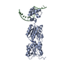 1jfsC  1jftC  1jh9C 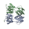 1dbqS C: citing same article ( S: Starting model for refinement |
|---|---|
| Similar structure data |

-Links
 PDBj
PDBj


- Assembly
| Deposited unit | 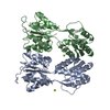
| ||||||||
|---|---|---|---|---|---|---|---|---|---|
| 1 |
| ||||||||
| Unit cell |
| ||||||||
| Details | The biological assembly of the PurR corepressor binding domain is a homodimer, which is contained within the asymmetric unit. |
-Components
| #1: Protein | Mass: 32451.217 Da / Num. of mol.: 2 Fragment: COREPRESSOR-FREE COREPRESSOR BINDING DOMAIN (Residues 53-341) Mutation: W147F Source method: isolated from a genetically manipulated source Source: (gene. exp.) Escherichia coli (E. coli) / Gene: purr / Plasmid: pET24a / Production host: Escherichia coli (E. coli) / Strain (production host): BL21 LAMBDA DE3 / References: UniProt: P0ACP7#2: Chemical |   Mass: 24.305 Da / Num. of mol.: 3 / Source method: obtained synthetically / Formula: Mg Mass: 24.305 Da / Num. of mol.: 3 / Source method: obtained synthetically / Formula: Mg#3: Water | ChemComp-HOH / | Water Mass: 18.015 Da / Num. of mol.: 150 / Source method: isolated from a natural source / Formula: H2O Mass: 18.015 Da / Num. of mol.: 150 / Source method: isolated from a natural source / Formula: H2O |
|---|
-Experimental details
-Experiment
| Experiment | Method: X-RAY DIFFRACTION / Number of used crystals: 1 |
|---|
- Sample preparation
| Crystal | Density Matthews: 2.25 Å3/Da / Density % sol: 45.35 % | |||||||||||||||||||||||||||||||||||
|---|---|---|---|---|---|---|---|---|---|---|---|---|---|---|---|---|---|---|---|---|---|---|---|---|---|---|---|---|---|---|---|---|---|---|---|---|
| Crystal grow | Temperature: 298 K / Method: vapor diffusion, hanging drop / pH: 7.6 Details: PEG 600, magnesium chloride, Tris HCl, pH 7.6, VAPOR DIFFUSION, HANGING DROP, temperature 298K | |||||||||||||||||||||||||||||||||||
| Crystal grow | *PLUS | |||||||||||||||||||||||||||||||||||
| Components of the solutions | *PLUS
|
-Data collection
| Diffraction | Mean temperature: 298 K |
|---|---|
| Diffraction source | Source: SYNCHROTRON / Site: SSRL  / Beamline: BL7-1 / Wavelength: 1.08 Å / Beamline: BL7-1 / Wavelength: 1.08 Å |
| Detector | Type: MARRESEARCH / Detector: IMAGE PLATE / Date: Jul 15, 1997 |
| Radiation | Monochromator: HORIZONTAL FOCUS Si(111) / Protocol: SINGLE WAVELENGTH / Monochromatic (M) / Laue (L): M / Scattering type: x-ray |
| Radiation wavelength | Wavelength: 1.08 Å / Relative weight: 1 |
| Reflection | Highest resolution: 2.4 Å / Num. all: 20934 / Num. obs: 20934 / % possible obs: 92.2 % / Observed criterion σ(F): 0 / Observed criterion σ(I): 0 / Redundancy: 3 % / Rmerge(I) obs: 0.079 / Net I/σ(I): 6.64 |
| Reflection shell | Resolution: 2.4→2.46 Å / Redundancy: 2.9 % / Rmerge(I) obs: 0.369 / Mean I/σ(I) obs: 1.9 / Num. unique all: 1562 / % possible all: 93.5 |
| Reflection | *PLUS |
| Reflection shell | *PLUS Lowest resolution: 2.45 Å / % possible obs: 93.5 % |
- Processing
| Software |
| ||||||||||||||||||||||||||||||||||||||||
|---|---|---|---|---|---|---|---|---|---|---|---|---|---|---|---|---|---|---|---|---|---|---|---|---|---|---|---|---|---|---|---|---|---|---|---|---|---|---|---|---|---|
| Refinement | Method to determine structure: MOLECULAR REPLACEMENT Starting model: 1DBQ Resolution: 2.4→10 Å / Isotropic thermal model: isotropic / σ(F): 0 / σ(I): 0 / Stereochemistry target values: Engh & Huber /
| ||||||||||||||||||||||||||||||||||||||||
| Displacement parameters | Biso mean: 2.662 Å2 | ||||||||||||||||||||||||||||||||||||||||
| Refinement step | Cycle: LAST / Resolution: 2.4→10 Å
| ||||||||||||||||||||||||||||||||||||||||
| Refine LS restraints |
| ||||||||||||||||||||||||||||||||||||||||
| Software | *PLUS Name: TNT / Classification: refinement | ||||||||||||||||||||||||||||||||||||||||
| Refinement | *PLUS Highest resolution: 2.4 Å | ||||||||||||||||||||||||||||||||||||||||
| Solvent computation | *PLUS | ||||||||||||||||||||||||||||||||||||||||
| Displacement parameters | *PLUS | ||||||||||||||||||||||||||||||||||||||||
| Refine LS restraints | *PLUS
|
